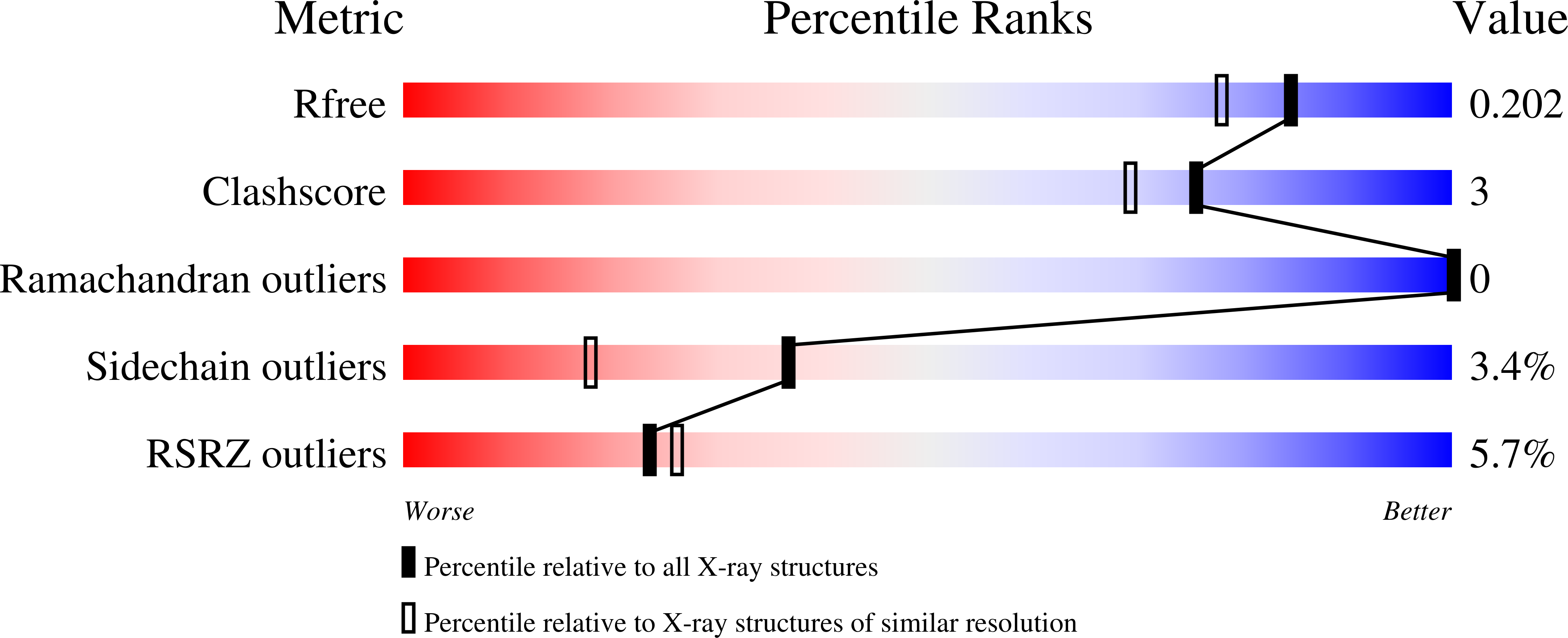
1RNJ image
Deposition Date 2003-12-01
Release Date 2004-09-07
Last Version Date 2023-08-23
Entry Detail
PDB ID:
1RNJ
Keywords:
Title:
Crystal structure of inactive mutant dUTPase complexed with substrate analogue imido-dUTP
Biological Source:
Source Organism:
Escherichia coli (Taxon ID: 562)
Method Details:
Experimental Method:
Resolution:
1.70 Å
R-Value Free:
0.18
R-Value Work:
0.15
R-Value Observed:
0.15
Space Group:
P 63 2 2
Macromolecular Entities
Polymer Type:polypeptide(L)
Molecule:Deoxyuridine 5'-triphosphate nucleotidohydrolase
Gene (Uniprot):dut
Mutations:D90N
Chain IDs:A
Chain Length:152
Number of Molecules:1
Biological Source:Escherichia coli
Primary Citation
Structural Insights into the Catalytic Mechanism of Phosphate Ester Hydrolysis by dUTPase.
J.Biol.Chem. 279 42907 42915 (2004)
PMID: 15208312 DOI: 10.1074/jbc.M406135200

Abstact

dUTPase is essential to keep uracil out of DNA. Crystal structures of substrate (dUTP and alpha,beta-imino-dUTP) and product complexes of wild type and mutant dUTPases were determined to reveal how an enzyme responsible for DNA integrity functions. A kinetic analysis of wild type and mutant dUTPases was performed to obtain relevant mechanistic information in solution. Substrate hydrolysis is shown to be initiated via in-line nucleophile attack of a water molecule oriented by an activating conserved aspartate residue. Substrate binding in a catalytically competent conformation is achieved by (i) multiple interactions of the triphosphate moiety with catalysis-assisting Mg2+, (ii) a concerted motion of residues from three conserved enzyme motifs as compared with the apoenzyme, and (iii) an intricate hydrogen-bonding network that includes several water molecules in the active site. Results provide an understanding for the catalytic role of conserved residues in dUTPases.

Legend

Protein

Chemical

Disease

Primary Citation of related structures