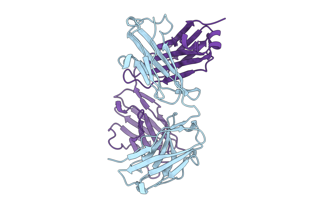
1RMF image
Deposition Date 1994-12-16
Release Date 1995-02-27
Last Version Date 2024-11-20
Entry Detail
PDB ID:
1RMF
Keywords:
Title:
STRUCTURES OF A MONOCLONAL ANTI-ICAM-1 ANTIBODY R6.5 FRAGMENT AT 2.8 ANGSTROMS RESOLUTION
Biological Source:
Source Organism:
Mus musculus (Taxon ID: 10090)
Method Details:
Experimental Method:
Resolution:
2.80 Å
R-Value Work:
0.18
R-Value Observed:
0.18
Space Group:
P 21 21 21
Macromolecular Entities
Polymer Type:polypeptide(L)
Molecule:IGG2A-KAPPA R6.5 FAB (HEAVY CHAIN)
Chain IDs:B (auth: H)
Chain Length:216
Number of Molecules:1
Biological Source:Mus musculus
Polymer Type:polypeptide(L)
Molecule:IGG2A-KAPPA R6.5 FAB (LIGHT CHAIN)
Chain IDs:A (auth: L)
Chain Length:219
Number of Molecules:1
Biological Source:Mus musculus
Primary Citation
Structure of a monoclonal anti-ICAM-1 antibody R6.5 Fab fragment at 2.8 A resolution.
Acta Crystallogr.,Sect.D 51 380 385 (1995)
PMID: 15299305 DOI: 10.1107/S0907444994011054

Abstact

The specific binding of the monoclonal murine anti-intercellular adhesion molecule-1 (anti-ICAM-1) antibody, R6.5, inhibits the attachment of neutrophils to endothelium and prevents the attachment of major group human rhinovirus (HRV) to ICAM-1. This binding interferes with the host immune system and, as a result, the R6.5 antibody has been developed as a therapeutic anti-inflammatory and perhaps anti-HRV agent. The variable-region amino-acid sequence of R6.5 was determined from the anti-ICAM-1 cDNA. The crystallization conditions of the Fab fragment of R6.5 were established and the three-dimensional structure was determined by X-ray crystallography. The crystal space group is orthorhombic P2(1)2(1)2(1), a = 40.36, b = 137.76, c = 91.32 A, and the highest resolution of recorded reflections is 2.7 A. The molecular-replacement method using known Fab structures was employed to solve the R6.5 Fab structure. The final R-factor is 18.8% for a total of 3320 non-H protein atoms, 39 water molecules and 10 606 unique reflections. The protein exhibits the typical immunoglobulin fold. The surface contour of the antigen-combining site of the R6.5 antibody has a wide groove which resembles more the structure of an anti-polypeptide antibody than the structure of an anti-protein antibody.

Legend

Protein

Chemical

Disease

Primary Citation of related structures