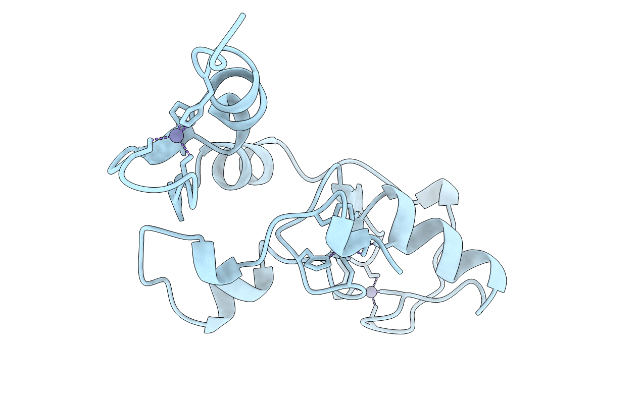
1RMD image
Deposition Date 1997-01-10
Release Date 1997-07-23
Last Version Date 2024-02-14
Entry Detail
PDB ID:
1RMD
Title:
RAG1 DIMERIZATION DOMAIN
Biological Source:
Source Organism:
Mus musculus (Taxon ID: 10090)
Host Organism:
Method Details:
Experimental Method:
Resolution:
2.10 Å
R-Value Free:
0.25
R-Value Work:
0.20
R-Value Observed:
0.20
Space Group:
P 32 2 1
Macromolecular Entities
Polymer Type:polypeptide(L)
Molecule:RAG1
Gene (Uniprot):Rag1
Chain IDs:A
Chain Length:116
Number of Molecules:1
Biological Source:Mus musculus
Ligand Molecules
Primary Citation
Crystal structure of the RAG1 dimerization domain reveals multiple zinc-binding motifs including a novel zinc binuclear cluster.
Nat.Struct.Biol. 4 586 591 (1997)
PMID: 9228952 DOI: 10.1038/nsb0797-586

Abstact

The crystal structure of the dimerization domain of the V(D)J recombination-activating protein, RAG1, was solved using zinc anomalous scattering. The structure reveals an unusual combination of multi-class zinc-binding motifs, including a zinc RING finger and a C2H2 zinc finger, that together from a single structural domain. The domain also contains a unique zinc binuclear cluster in place of a normally mononuclear zinc site in the RING finger. Together, four zinc ions help organize the entire domain, including the two helices that form the dimer interface.

Legend

Protein

Chemical

Disease

Primary Citation of related structures