
Deposition Date
1997-10-28
Release Date
1998-02-25
Last Version Date
2024-02-14
Entry Detail
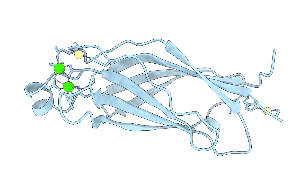
PDB ID:
1RLW
Keywords:
Title:
CALCIUM-PHOSPHOLIPID BINDING DOMAIN FROM CYTOSOLIC PHOSPHOLIPASE A2
Biological Source:
Source Organism(s):
Homo sapiens (Taxon ID: 9606)
Expression System(s):
Method Details:
Experimental Method:
Resolution:
2.40 Å
R-Value Free:
0.27
R-Value Work:
0.22
Space Group:
P 31 2 1
