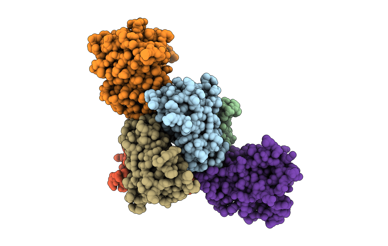
1RLB image
Deposition Date 1995-02-20
Release Date 1996-04-11
Last Version Date 2024-10-16
Entry Detail
PDB ID:
1RLB
Title:
RETINOL BINDING PROTEIN COMPLEXED WITH TRANSTHYRETIN
Biological Source:
Source Organism:
Homo sapiens (Taxon ID: 9606)
Gallus gallus (Taxon ID: 9031)
Method Details:
Experimental Method:
Resolution:
3.10 Å
R-Value Work:
0.21
R-Value Observed:
0.21
Space Group:
I 2 2 2
Macromolecular Entities
Polymer Type:polypeptide(L)
Molecule:TRANSTHYRETIN
Gene (Uniprot):TTR
Chain IDs:A, B, C, D
Chain Length:127
Number of Molecules:4
Biological Source:Homo sapiens
Polymer Type:polypeptide(L)
Molecule:RETINOL BINDING PROTEIN
Gene (Uniprot):RBP4
Chain IDs:E, F
Chain Length:174
Number of Molecules:2
Biological Source:Gallus gallus
Ligand Molecules
Primary Citation
Structure of a complex of two plasma proteins: transthyretin and retinol-binding protein.
Science 268 1039 1041 (1995)
PMID: 7754382

Abstact

The three-dimensional structure of the complex formed by two plasma proteins, transthyretin and retinol-binding protein, was determined from x-ray diffraction data to a nominal resolution of 3.1 angstroms. One tetramer of transthyretin was bound to two molecules of retinol-binding protein. The two retinol-binding protein molecules established molecular interactions with the same transthyretin dimer, and each also made contacts with one of the other two monomers. Thus, the other two potential binding sites in a transthyretin tetramer were blocked. The amino acid residues of the retinol-binding protein that were involved in the contacts were close to the retinol-binding site.

Legend

Protein

Chemical

Disease

Primary Citation of related structures