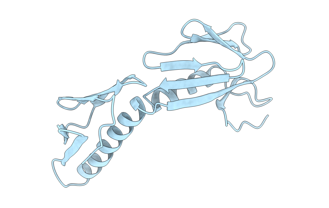
1RL6 image
Deposition Date 1999-01-14
Release Date 1999-02-02
Last Version Date 2023-12-27
Entry Detail
PDB ID:
1RL6
Title:
RIBOSOMAL PROTEIN L6
Biological Source:
Source Organism:
Method Details:
Experimental Method:
Resolution:
2.00 Å
R-Value Free:
0.30
R-Value Work:
0.25
Space Group:
P 61 2 2
Macromolecular Entities
Polymer Type:polypeptide(L)
Molecule:PROTEIN (RIBOSOMAL PROTEIN L6)
Gene (Uniprot):rplF
Chain IDs:A
Chain Length:177
Number of Molecules:1
Biological Source:Geobacillus stearothermophilus
Primary Citation
Ribosomal protein L6: structural evidence of gene duplication from a primitive RNA binding protein.
EMBO J. 12 4901 4908 (1993)
PMID: 8262035

Abstact

In all cells, protein synthesis is coordinated by the ribosome, a large ribonucleoprotein particle that is composed of > 50 distinct protein molecules and several large RNA molecules. Here we present the crystal structure of ribosomal protein L6 from the thermophilic bacterium Bacillus stearothermophilus solved at 2.6 A resolution. L6 contains two domains with almost identical folds, implying that it was created by an ancient gene duplication event. The surface of the molecule displays several likely sites of interaction with other components of the ribosome. The RNA binding sites appear to be localized in the C-terminal domain whereas the N-terminal domain contains the potential sites for protein-protein interactions. The domain structure is homologous with several other ribosomal proteins and to a large family of eukaryotic RNA binding proteins.

Legend

Protein

Chemical

Disease

Primary Citation of related structures