1RL0 image
Deposition Date 2003-11-24
Release Date 2004-12-07
Last Version Date 2023-08-23
Entry Detail
PDB ID:
1RL0
Title:
Crystal structure of a new ribosome-inactivating protein (RIP): dianthin 30
Biological Source:
Source Organism:
Method Details:
Experimental Method:
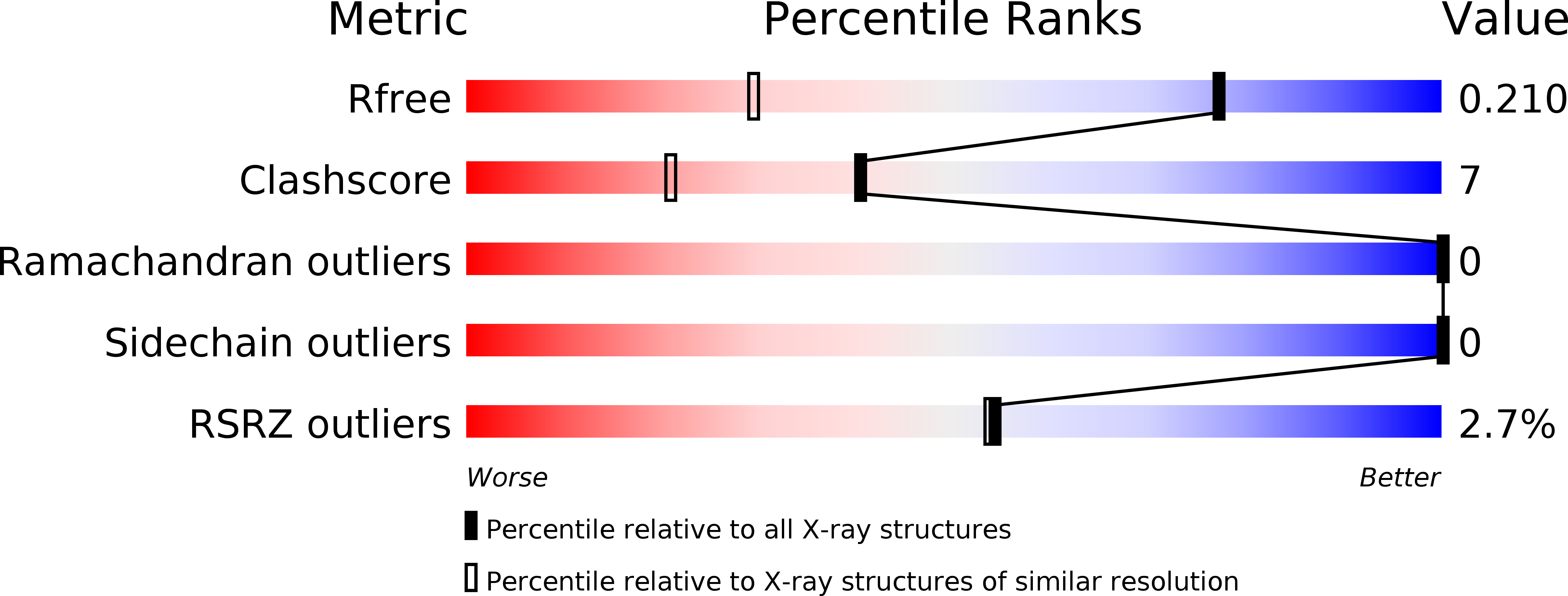
Resolution:
1.40 Å
R-Value Free:
0.20
R-Value Work:
0.18
R-Value Observed:
0.18
Space Group:
P 1 21 1
Macromolecular Entities
Structures with similar UniProt ID
Protein Blast
Polymer Type:polypeptide(L)
Molecule:Antiviral protein DAP-30
Chain IDs:A
Chain Length:255
Number of Molecules:1
Biological Source:Dianthus caryophyllus
Primary Citation
The 1.4A structure of dianthin 30 indicates a role of surface potential at the active site of type 1 ribosome inactivating proteins
J.Struct.Biol. 149 204 212 (2005)
PMID: 15681236 DOI: 10.1016/j.jsb.2004.11.007

Abstact

Ribosome inactivating proteins (RIPs) are plant proteins with enzymatic activity identified as rRNA N-glycosidase (EC 3.2.2.22), which cleaves the N-glycosidic bond of a specific adenine on the ricin/sarcin region of rRNA, thus causing inhibition of protein synthesis. They also depurinate extensively DNA and other polynucleotides. The three-dimensional structure of dianthin 30, a type 1 (single-chain) RIP of Dianthus caryophyllus (leaves), is now described at 1.4 angstroms, a resolution never achieved before for any RIP. The fold typical of RIPs is conserved, despite some differences in the loop regions. The general structure comparison by superimposed alpha-carbon (249 atoms) and the sequence alignment by structure for dianthin 30 and saporin-S6 give a root mean square deviation of 0.625 angstroms. Despite the differences reported for the biological activities of the two RIPs, their structures fit quite well and both show a protein segment containing strands beta7, beta8, and beta9 shorter than other RIPs. However, the surface electrostatic potential in the active site region neatly distinguishes dianthin 30 from saporin-S6. The possible relationship between the charge distribution and the behavior of the proteins toward different substrates is discussed.

Legend

Protein

Chemical

Disease

Primary Citation of related structures
Feedback Form
Name
Email
Institute
Feedback