Deposition Date
1999-05-20
Release Date
1999-09-01
Last Version Date
2023-08-23
Entry Detail
PDB ID:
1RK2
Keywords:
Title:
E. COLI RIBOKINASE COMPLEXED WITH RIBOSE AND ADP, SOLVED IN SPACE GROUP P212121
Biological Source:
Source Organism(s):
Escherichia coli (Taxon ID: 562)
Expression System(s):
Method Details:
Experimental Method:
Resolution:
2.25 Å
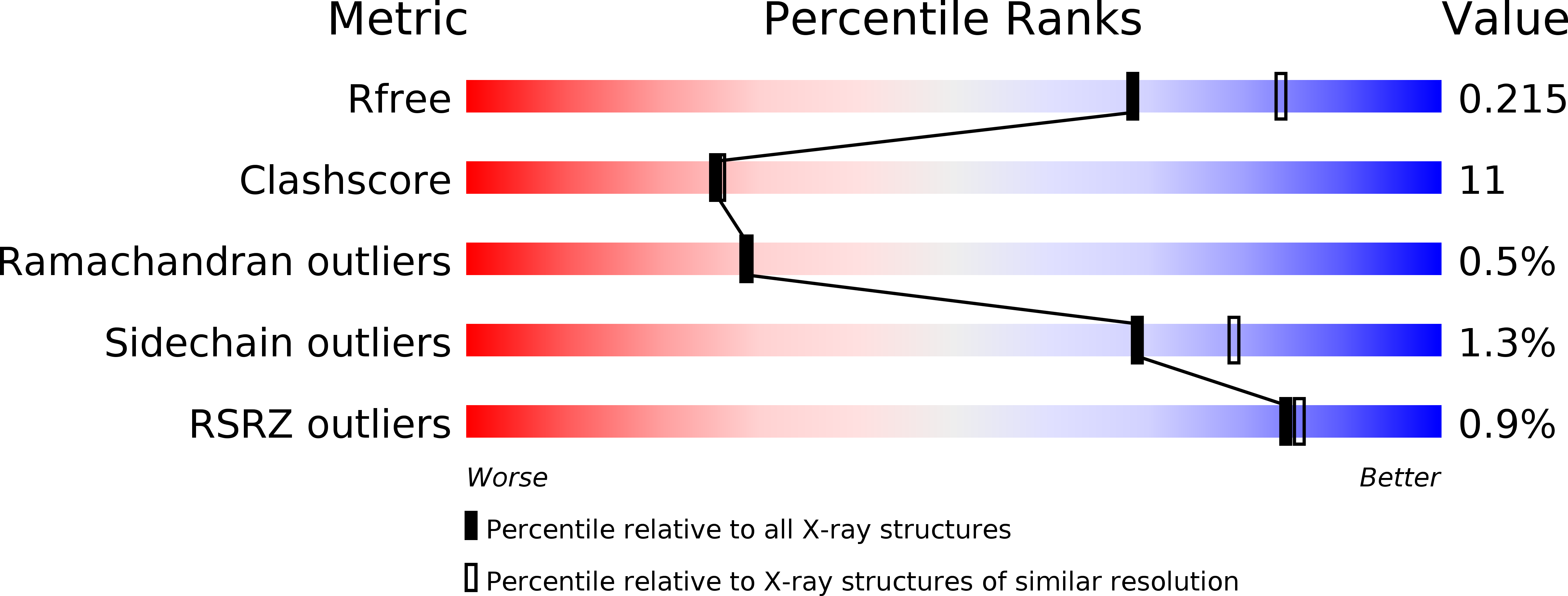
R-Value Free:
0.26
R-Value Work:
0.22
Space Group:
P 21 21 21