1RJU image
Deposition Date 2003-11-20
Release Date 2004-12-07
Last Version Date 2024-02-14
Entry Detail
PDB ID:
1RJU
Title:
Crystal structure of a truncated form of yeast copper thionein
Biological Source:
Source Organism:
Method Details:
Experimental Method:
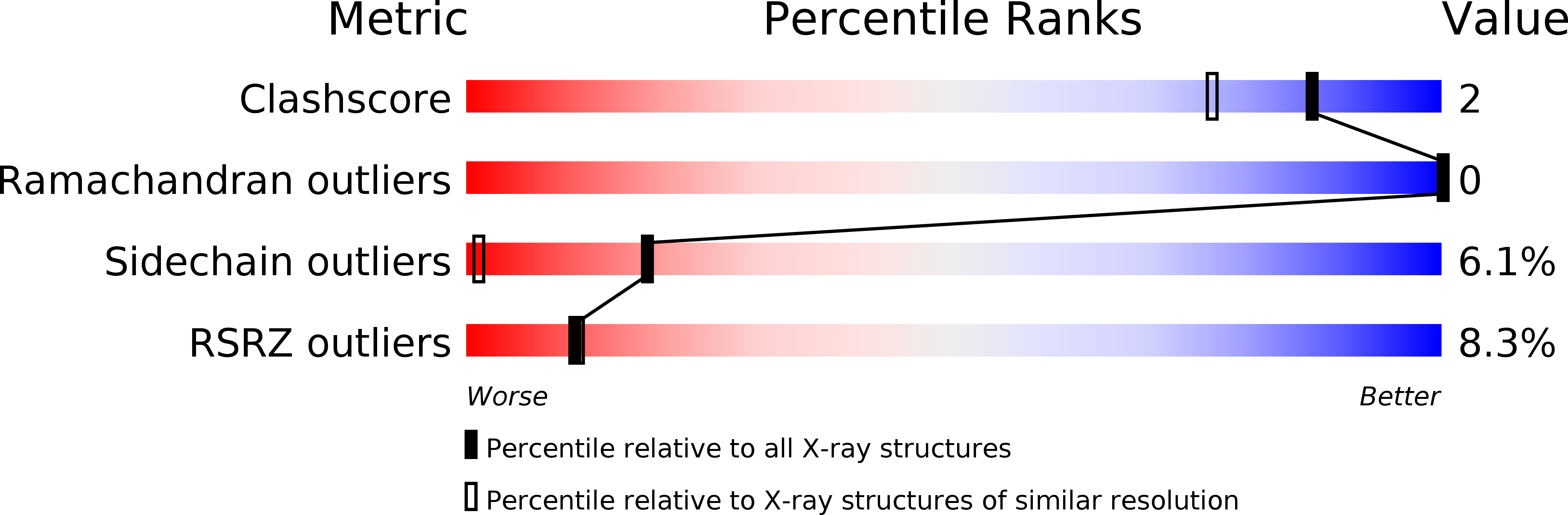
Resolution:
1.44 Å
R-Value Free:
0.17
R-Value Work:
0.14
R-Value Observed:
0.14
Space Group:
P 43 3 2
Macromolecular Entities
Structures with similar UniProt ID
Protein Blast
Polymer Type:polypeptide(L)
Molecule:Metallothionein
Chain IDs:A (auth: V)
Chain Length:36
Number of Molecules:1
Biological Source:
Ligand Molecules
Primary Citation
The Crystal Structure of Yeast Copper Thionein: the solution of a long lasting Enigma.
Proc.Natl.Acad.Sci.USA 102 51 56 (2005)
PMID: 15613489 DOI: 10.1073/pnas.0408254101

Abstact

We report here the crystal structure of yeast copper thionein (Cu-MT), determined at 1.44-A resolution. The Cu-MT structure shows the largest known oligonuclear Cu(I) thiolate cluster in biology, consisting of six trigonally and two digonally coordinated Cu(I) ions. This is at variance with the results from previous spectroscopic determinations, which were performed on MT samples containing seven rather than eight metal ions. The protein backbone has a random coil structure with the loops enfolding the copper cluster, which is located in a cleft where it is bound to 10 cysteine residues. The protein structure is somewhat different from that of Ag(7)-MT and similar, but not identical, to that of Cu(7)-MT. Besides the different structure of the metal cluster, the main differences lie in the cysteine topology and in the conformation of some portions of the backbone. The present structure suggests that Cu-MT, in addition to its role as a safe depository for copper ions in the cell, may play an active role in the delivery of copper to metal-free chaperones.

Legend

Protein

Chemical

Disease

Primary Citation of related structures