Deposition Date
2003-11-18
Release Date
2004-07-20
Last Version Date
2024-05-01
Entry Detail
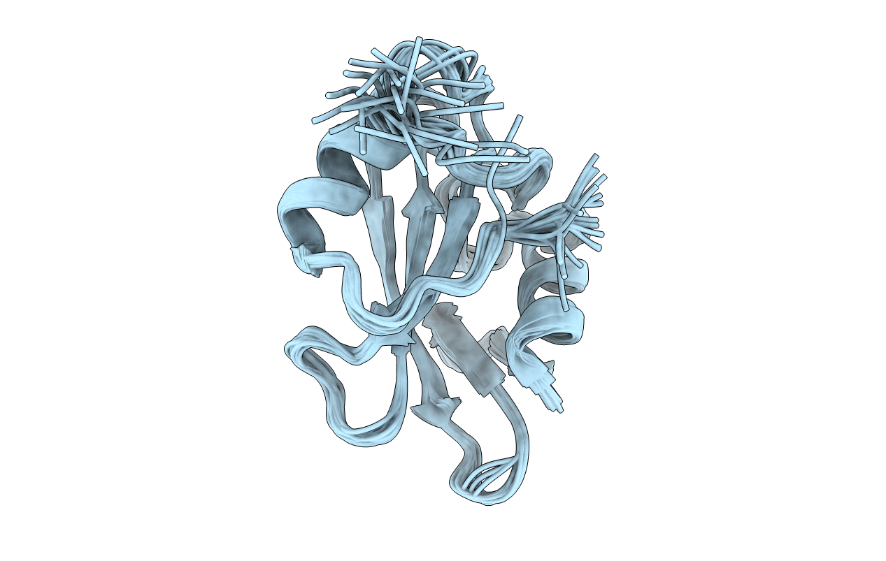
PDB ID:
1RJA
Keywords:
Title:
Solution Structure and Backbone Dynamics of the Nonreceptor Tyrosine Kinase PTK6/Brk SH2 Domain
Biological Source:
Source Organism(s):
Homo sapiens (Taxon ID: 9606)
Expression System(s):
Method Details:
Experimental Method:
Conformers Calculated:
50
Conformers Submitted:
21
Selection Criteria:
structures with the lowest energy