1RJ8 image
Deposition Date 2003-11-18
Release Date 2003-12-09
Last Version Date 2024-11-13
Entry Detail
PDB ID:
1RJ8
Title:
The crystal structure of TNF family member EDA-A2
Biological Source:
Source Organism:
Homo sapiens (Taxon ID: 9606)
Method Details:
Experimental Method:
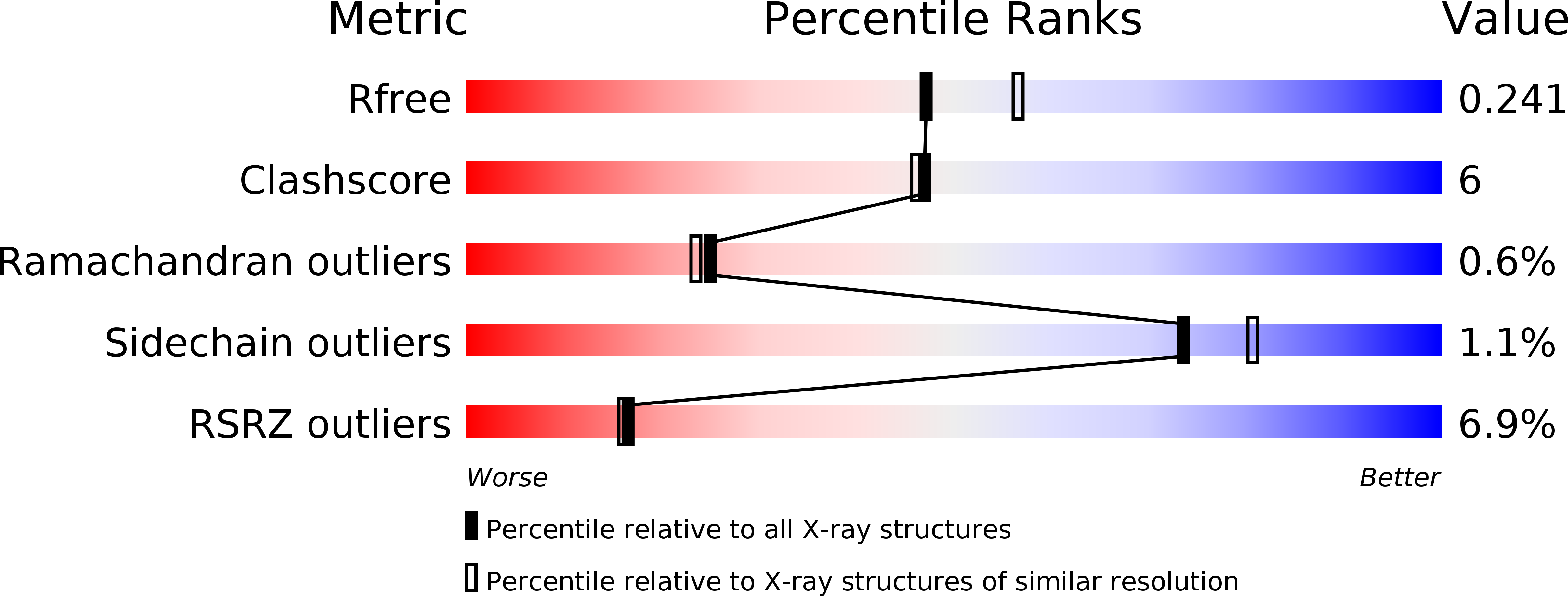
Resolution:
2.23 Å
R-Value Free:
0.24
R-Value Work:
0.20
R-Value Observed:
0.20
Space Group:
P 1 21 1
Macromolecular Entities
Structures with similar UniProt ID
Protein Blast
Polymer Type:polypeptide(L)
Molecule:ectodysplasin-A isoform EDA-A2
Gene (Uniprot):EDA
Chain IDs:A, B, C (auth: D), D (auth: E), E (auth: F), F (auth: G)
Chain Length:164
Number of Molecules:6
Biological Source:Homo sapiens
Primary Citation
The Crystal Structure of EDA-A1 and EDA-A2: splice variants with distinct receptor specificity
STRUCTURE 11 1513 1520 (2003)
PMID: 14656435 DOI: 10.1016/j.str.2003.11.009

Abstact

EDA is a tumor necrosis factor family member involved in ectodermal development. Splice variants EDA-A1 and EDA-A2 differ only by the presence of Glu 308 and Val 309 in the expected receptor binding region of EDA-A1 but not EDA-A2. This two amino acid difference functions as a switch controlling receptor specificity. EDA-A1 binds only to EDAR, while EDA-A2 is specific for XEDAR. In order to understand the structural basis of this switch, we determined the X-ray crystal structures of the TNF domain of both EDA-A1 and EDA-A2 at 2.3 A and 2.2 A, respectively. While the backbone conformation around the splice difference is similar in both isoforms, the conformation of the following loop, the surface charge, and the shape of the expected receptor binding site differ significantly.

Legend

Protein

Chemical

Disease

Primary Citation of related structures