1RJ6 image
Deposition Date 2003-11-18
Release Date 2004-03-09
Last Version Date 2024-10-30
Entry Detail
PDB ID:
1RJ6
Keywords:
Title:
Crystal Structure of the Extracellular Domain of Murine Carbonic Anhydrase XIV in Complex with Acetazolamide
Biological Source:
Source Organism:
Mus musculus (Taxon ID: 10090)
Host Organism:
Method Details:
Experimental Method:
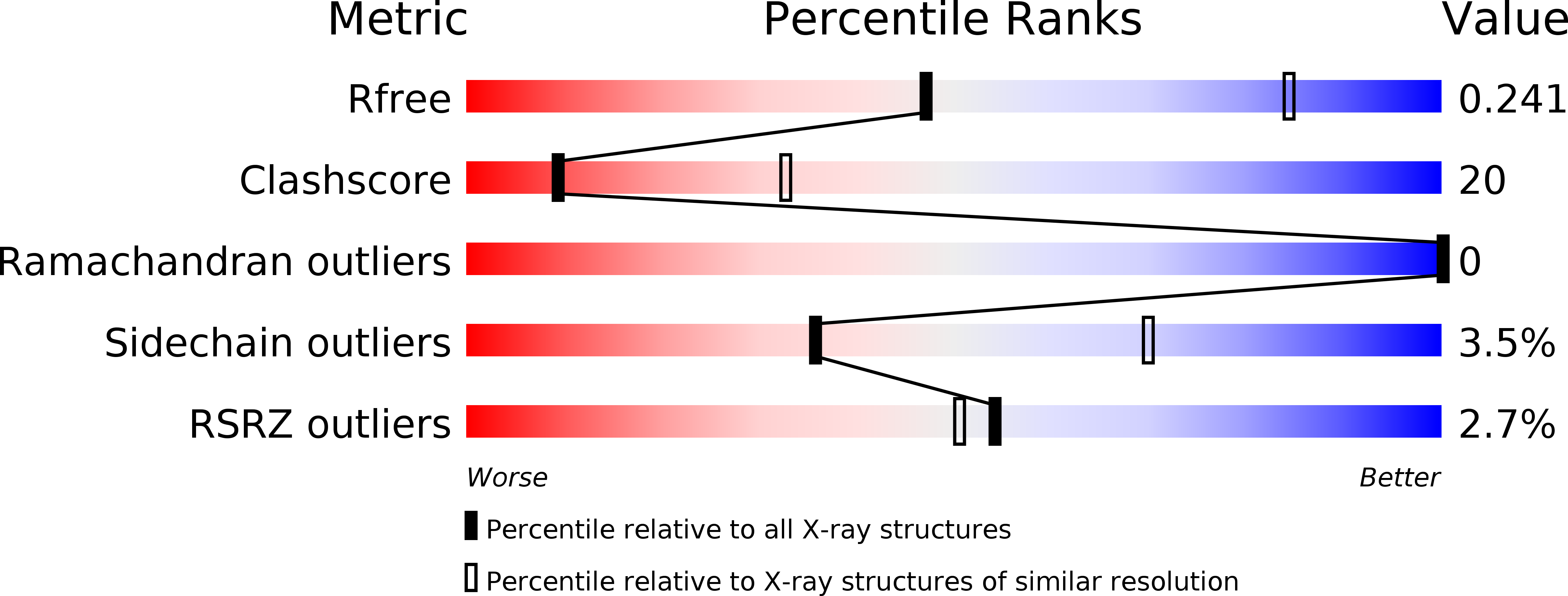
Resolution:
2.90 Å
R-Value Free:
0.25
R-Value Work:
0.20
R-Value Observed:
0.20
Space Group:
P 1 21 1
Macromolecular Entities
Structures with similar UniProt ID
Protein Blast
Polymer Type:polypeptide(L)
Molecule:Carbonic anhydrase XIV
Gene (Uniprot):Ca14
Chain IDs:A, B
Chain Length:261
Number of Molecules:2
Biological Source:Mus musculus
Modified Residue
Compound ID Chain ID Parent Comp ID Details 2D Image
ASN A ASN GLYCOSYLATION SITE
Primary Citation
Expression, assay, and structure of the extracellular domain of murine carbonic anhydrase XIV: implications for selective inhibition of membrane-associated isozymes.
J.Biol.Chem. 279 7223 7228 (2004)
PMID: 14660577 DOI: 10.1074/jbc.M310809200

Abstact

Carbonic anhydrase (CA) XIV is the most recently identified mammalian carbonic anhydrase isozyme, and its presence has been demonstrated in a number of tissues. Full-length CA XIV is a transmembrane protein composed of an extracellular catalytic domain, a single transmembrane helix, and a short intracellular polypeptide segment. The amino acid sequence identity of human CA XIV relative to the other membrane-associated isozymes (CA IV, CA IX, and CA XII) is 34-46%. We report here the expression and purification of both the full-length enzyme and a truncated, secretory form of murine CA XIV. Both forms of this isozyme are highly active, and both show an abrogation of activity in the presence of 0.2% SDS, in contrast to the behavior of murine CA IV. We also report the crystal structure of the extracellular domain of murine CA XIV at 2.8 A resolution and of an enzyme-acetazolamide complex at 2.9 A resolution. The structure shows a monomeric glycoprotein with a topology similar to that of other mammalian CA isozymes. Based on the x-ray crystallographic results, we compare and contrast known structures of membrane-associated CA isozymes to rationalize the structural elements responsible for the SDS resistance of CA IV and to discuss prospects for the design of selective inhibitors of membrane-associated CA isozymes.

Legend

Protein

Chemical

Disease

Primary Citation of related structures
Feedback Form
Name
Email
Institute
Feedback