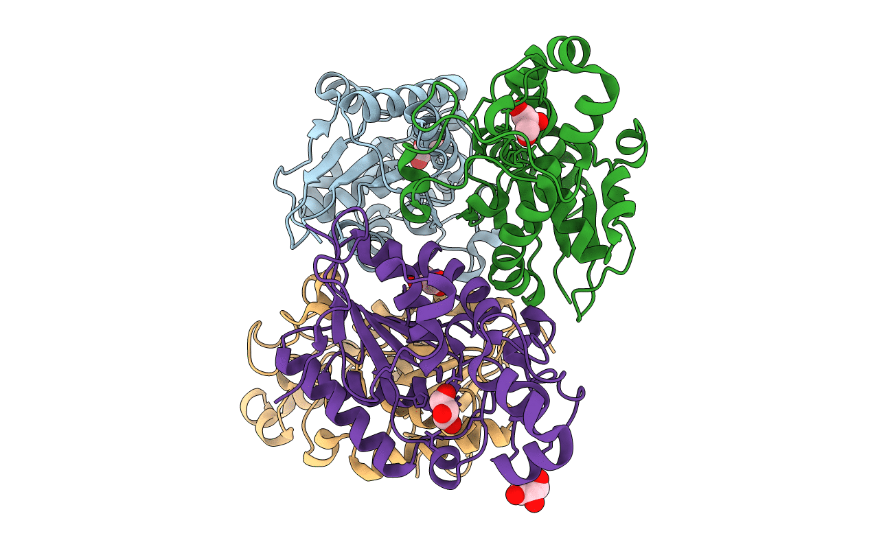
1RII image
Deposition Date 2003-11-17
Release Date 2004-10-05
Last Version Date 2023-08-23
Entry Detail
PDB ID:
1RII
Keywords:
Title:
Crystal structure of phosphoglycerate mutase from M. Tuberculosis
Biological Source:
Source Organism:
Host Organism:
Method Details:
Experimental Method:
Resolution:
1.70 Å
R-Value Free:
0.26
R-Value Observed:
0.21
Space Group:
P 1 21 1
Macromolecular Entities
Polymer Type:polypeptide(L)
Molecule:2,3-bisphosphoglycerate-dependent phosphoglycerate mutase
Chain IDs:A, B, C, D
Chain Length:265
Number of Molecules:4
Biological Source:Mycobacterium tuberculosis
Ligand Molecules
Primary Citation
The 1.70 angstroms X-ray crystal structure of Mycobacterium tuberculosis phosphoglycerate mutase.
Acta Crystallogr.,Sect.D 61 309 315 (2005)
PMID: 15735341 DOI: 10.1107/S0907444904033190

Abstact

The single-crystal X-ray structure of phosphoglycerate mutase from Mycobacterium tuberculosis has been determined at a resolution of 1.70 angstroms. The C-terminal tail of each of the subunits is flexible and disordered; however, for one of the four chains (chain A) all but five residues of the chain could be modeled. Noteworthy features of the structure include the active site and a proline-rich segment in each monomer forming a short left-handed polyprolyl helix. These segments lie on the enzyme surface and could conceivably participate in protein-protein interactions.

Legend

Protein

Chemical

Disease

Primary Citation of related structures