1RIH image
Deposition Date 2003-11-17
Release Date 2004-01-13
Last Version Date 2024-10-30
Entry Detail
PDB ID:
1RIH
Keywords:
Title:
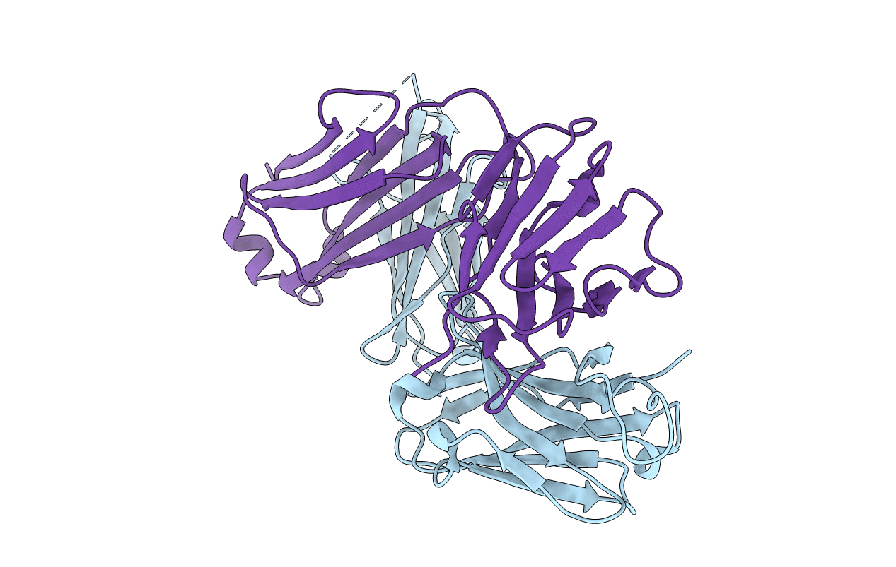
Crystal Structure of Fab 14F7, a unique anti-tumor antibody specific for N-glycolyl GM3
Biological Source:
Source Organism:
Mus musculus (Taxon ID: 10090)
Method Details:
Experimental Method:
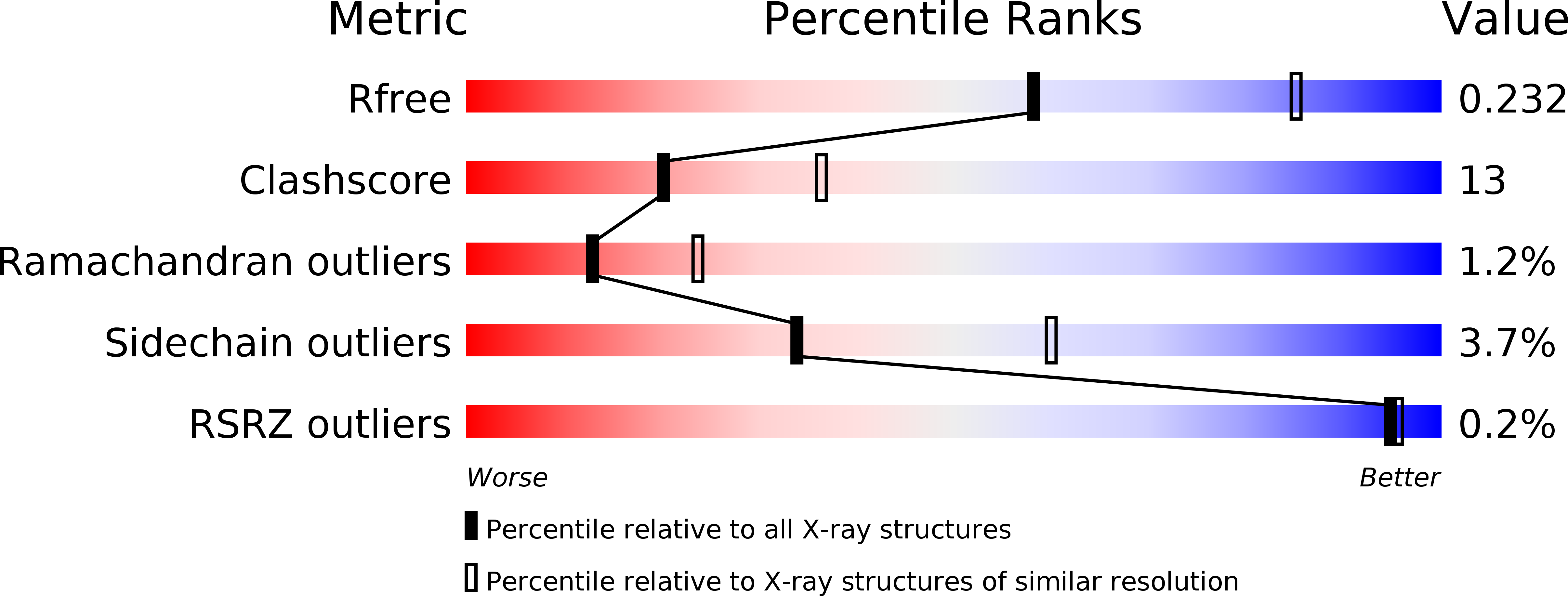
Resolution:
2.50 Å
R-Value Free:
0.23
R-Value Work:
0.18
R-Value Observed:
0.18
Space Group:
P 21 21 21
Macromolecular Entities
Structures with similar UniProt ID
Protein Blast
Polymer Type:polypeptide(L)
Molecule:heavy chain of antibody 14F7
Chain IDs:B (auth: H)
Chain Length:231
Number of Molecules:1
Biological Source:Mus musculus
Structures with similar UniProt ID
Protein Blast
Polymer Type:polypeptide(L)
Molecule:light chain of antibody 14F7
Chain IDs:A (auth: L)
Chain Length:214
Number of Molecules:1
Biological Source:Mus musculus
Primary Citation
Structure and Molecular Interactions of a Unique Antitumor Antibody Specific for N-Glycolyl GM3.
J.Biol.Chem. 279 5597 5603 (2004)
PMID: 14627696 DOI: 10.1074/jbc.M311693200

Abstact

N-glycolyl GM3 ganglioside is an attractive target antigen for cancer immunotherapy, because this epitope is a molecular marker of certain tumor cells and not expressed in normal human tissues. The murine monoclonal antibody 14F7 specifically recognizes N-glycolyl GM3 and shows no cross-reactivity with the abundant N-acetyl GM3 ganglioside, a close structural homologue of N-glycolyl GM3. Here, we report the crystal structure of the 14F7 Fab fragment at 2.5 A resolution and its molecular model with the saccharide moiety of N-glycolyl GM3, NeuGcalpha3Galbeta4Glcbeta. Fab 14F7 contains a very long CDR H3 loop, which divides the antigen-binding site of this antibody into two subsites. In the docking model, the saccharide ligand is bound to one of these subsites, formed solely by heavy chain residues. The discriminative feature of N-glycolyl GM3 versus N-acetyl GM3, its hydroxymethyl group, is positioned in a hydrophilic cavity, forming hydrogen bonds with the carboxyl group of Asp H52, the indole NH of Trp H33 and the hydroxyl group of Tyr H50. For the hydrophobic methyl group of N-acetyl GM3, this environment would not be favorable, explaining why the antibody specifically recognizes N-glycolyl GM3, but not N-acetyl GM3. Mutation of Asp H52 to hydrophobic residues of similar size completely abolished binding. Our model of the antibodycarbohydrate complex is consistent with binding data for several tested glycolipids as well as for a variety of 14F7 mutants with replaced VL domains.

Legend

Protein

Chemical

Disease

Primary Citation of related structures
Feedback Form
Name
Email
Institute
Feedback