
Deposition Date
2003-11-13
Release Date
2004-03-09
Last Version Date
2024-02-14
Entry Detail
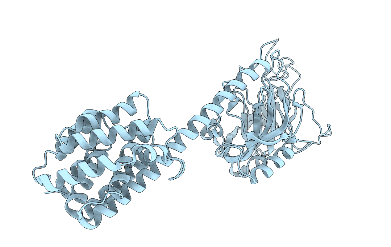
PDB ID:
1RH1
Keywords:
Title:
crystal structure of the cytotoxic bacterial protein colicin B at 2.5 A resolution
Biological Source:
Source Organism(s):
Escherichia coli (Taxon ID: 562)
Expression System(s):
Method Details:
Experimental Method:
Resolution:
2.50 Å
R-Value Free:
0.24
R-Value Work:
0.19
R-Value Observed:
0.22
Space Group:
C 2 2 21
