Deposition Date
2003-11-12
Release Date
2004-07-27
Last Version Date
2023-08-23
Entry Detail
PDB ID:
1RGI
Keywords:
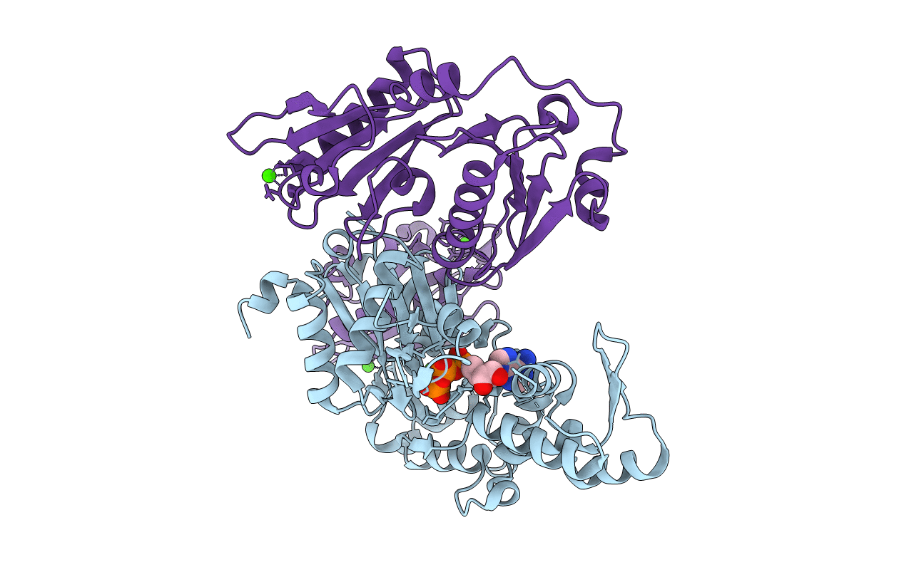
Title:
Crystal structure of gelsolin domains G1-G3 bound to actin
Biological Source:
Source Organism(s):
Equus caballus (Taxon ID: 9796)
Oryctolagus cuniculus (Taxon ID: 9986)
Oryctolagus cuniculus (Taxon ID: 9986)
Method Details:
Experimental Method:
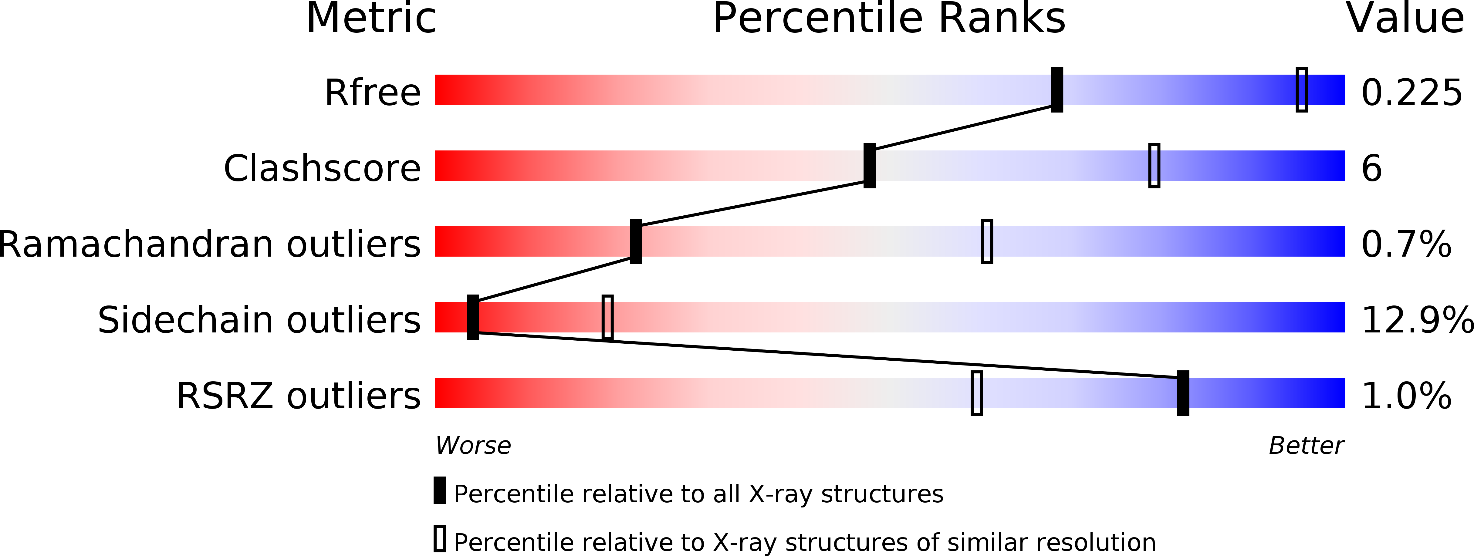
Resolution:
3.00 Å
R-Value Free:
0.25
R-Value Work:
0.22
R-Value Observed:
0.22
Space Group:
P 31 2 1