1RG8 image
Deposition Date 2003-11-11
Release Date 2004-10-05
Last Version Date 2023-08-23
Entry Detail
PDB ID:
1RG8
Title:
Human Acidic Fibroblast Growth Factor (haFGF-1) at 1.10 angstrom resolution (140 amino acid form)
Biological Source:
Source Organism:
Homo sapiens (Taxon ID: 9606)
Host Organism:
Method Details:
Experimental Method:
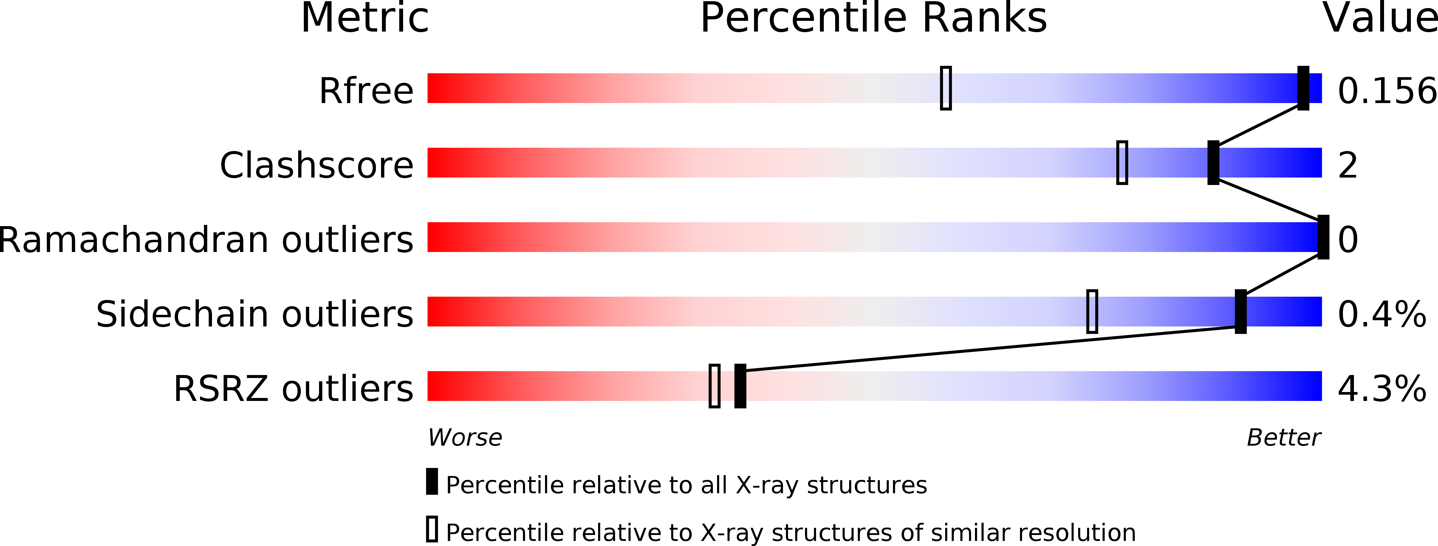
Resolution:
1.10 Å
R-Value Free:
0.17
R-Value Work:
0.14
R-Value Observed:
0.14
Space Group:
C 2 2 21
Macromolecular Entities
Polymer Type:polypeptide(L)
Molecule:Heparin-binding growth factor 1
Gene (Uniprot):FGF1
Chain IDs:A, B
Chain Length:146
Number of Molecules:2
Biological Source:Homo sapiens
Ligand Molecules
Primary Citation
An atomic resolution structure for human fibroblast growth factor 1.
Proteins 57 626 634 (2004)
PMID: 15382229 DOI: 10.1002/prot.20239

Abstact

A 1.10-A atomic resolution X-ray structure of human fibroblast growth factor 1 (FGF-1), a member of the beta-trefoil superfold, has been determined. The beta-trefoil is one of 10 fundamental protein superfolds and is the only superfold to exhibit 3-fold structural symmetry (comprising 3 "trefoil" units). The quality of the diffraction data permits unambiguous assignment of Asn, Gln, and His rotamers, Pro ring pucker, as well as refinement of atomic anisotropic displacement parameters (ADPs). The FGF-1 structure exhibits numerous core-packing defects, detectable using a 1.0-A probe radius. In addition to contributing to the relatively low thermal stability of FGF-1, these defects may also permit domain motions within the structure. The availability of refined ADPs allows a translation/libration/screw (TLS) analysis of putative rigid body domains. The TLS analysis shows that beta-strands 6-12 together form a rigid body, and there is a clear demarcation in TLS motions between the adjacent carboxyl- and amino-termini. Although separate from beta-strands 6-12, the individual beta-strands 1-5 do not exhibit correlated motions; thus, this region appears to be comparatively flexible. The heparin-binding contacts of FGF-1 are located within beta-strands 6-12; conversely, a significant portion of the receptor-binding contacts are located within beta-strands 1-5. Thus, the observed rigid body motion in FGF-1 appears related to the ligand-binding functionalities.

Legend

Protein

Chemical

Disease

Primary Citation of related structures