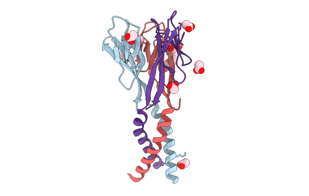
1RFX image
Deposition Date 2003-11-10
Release Date 2004-06-08
Last Version Date 2024-11-13
Entry Detail
PDB ID:
1RFX
Title:
Crystal Structure of resisitin
Biological Source:
Source Organism:
Mus musculus (Taxon ID: 10090)
Host Organism:
Method Details:
Experimental Method:
Resolution:
2.00 Å
R-Value Free:
0.21
R-Value Work:
0.17
R-Value Observed:
0.17
Space Group:
C 2 2 21
Macromolecular Entities
Polymer Type:polypeptide(L)
Molecule:Resistin
Gene (Uniprot):Retn
Chain IDs:A, B, C
Chain Length:94
Number of Molecules:3
Biological Source:Mus musculus
Primary Citation
Disulfide-dependent multimeric assembly of resistin family hormones
Science 304 1154 1158 (2004)
PMID: 15155948 DOI: 10.1126/science.1093466

Abstact

Resistin, founding member of the resistin-like molecule (RELM) hormone family, is secreted selectively from adipocytes and induces liver-specific antagonism of insulin action, thus providing a potential molecular link between obesity and diabetes. Crystal structures of resistin and RELMbeta reveal an unusual multimeric structure. Each protomer comprises a carboxy-terminal disulfide-rich beta-sandwich "head" domain and an amino-terminal alpha-helical "tail" segment. The alpha-helical segments associate to form three-stranded coiled coils, and surface-exposed interchain disulfide linkages mediate the formation of tail-to-tail hexamers. Analysis of serum samples shows that resistin circulates in two distinct assembly states, likely corresponding to hexamers and trimers. Infusion of a resistin mutant, lacking the intertrimer disulfide bonds, in pancreatic-insulin clamp studies reveals substantially more potent effects on hepatic insulin sensitivity than those observed with wild-type resistin. This result suggests that processing of the intertrimer disulfide bonds may reflect an obligatory step toward activation.

Legend

Protein

Chemical

Disease

Primary Citation of related structures