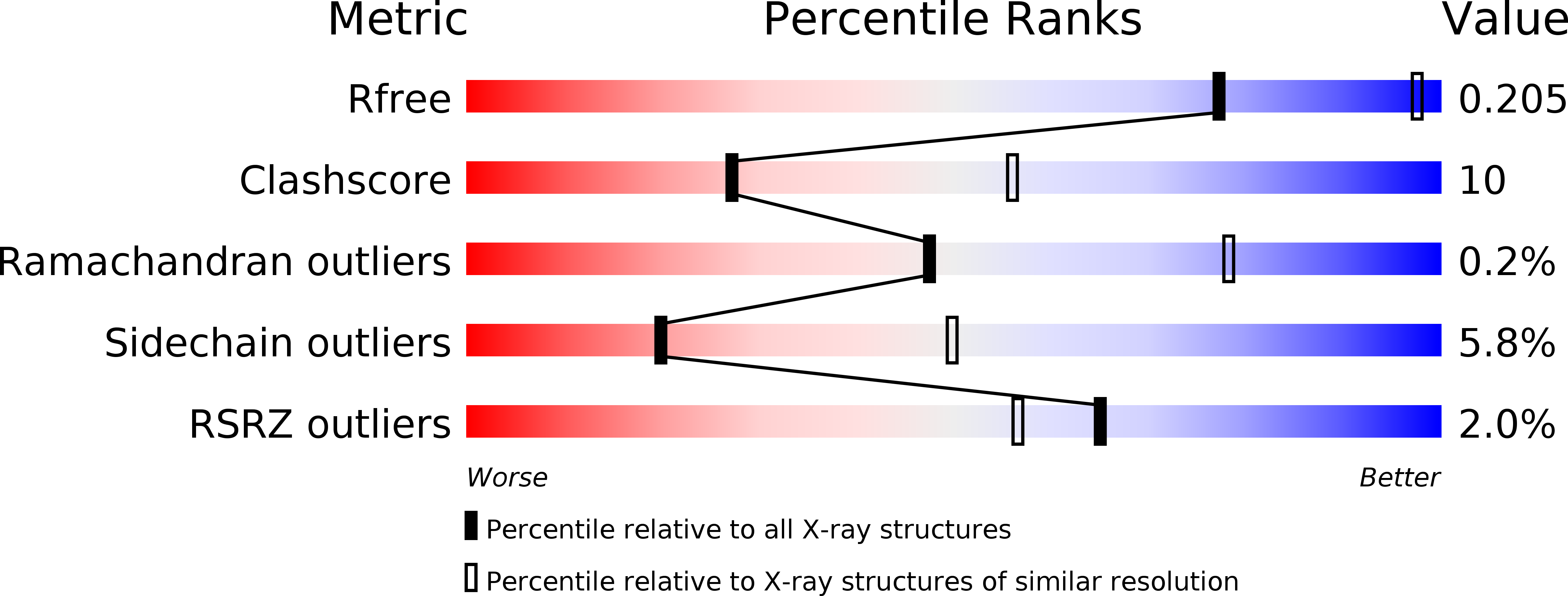
1RFV image
Deposition Date 2003-11-10
Release Date 2004-04-27
Last Version Date 2023-10-25
Entry Detail
PDB ID:
1RFV
Keywords:
Title:
Crystal structure of pyridoxal kinase complexed with ADP
Biological Source:
Source Organism:
Ovis aries (Taxon ID: 9940)
Method Details:
Experimental Method:
Resolution:
2.80 Å
R-Value Free:
0.22
R-Value Work:
0.19
R-Value Observed:
0.19
Space Group:
P 21 21 21
Macromolecular Entities
Polymer Type:polypeptide(L)
Molecule:pyridoxal kinase
Gene (Uniprot):PDXK
Chain IDs:A, B
Chain Length:312
Number of Molecules:2
Biological Source:Ovis aries
Primary Citation

Abstact

To understand the processes involved in the catalytic mechanism of pyridoxal kinase (PLK),1 we determined the crystal structures of PLK.AMP-PCP-pyridoxamine, PLK.ADP.PLP, and PLK.ADP complexes. Comparisons of these structures have revealed that PLK exhibits different conformations during its catalytic process. After the binding of AMP-PCP (an analogue that replaced ATP) and pyridoxamine to PLK, this enzyme retains a conformation similar to that of the PLK.ATP complex. The distance between the reacting groups of the two substrates is 5.8 A apart, indicating that the position of ATP is not favorable to spontaneous transfer of its phosphate group. However, the structure of PLK.ADP.PLP complex exhibited significant changes in both the conformation of the enzyme and the location of the ligands at the active site. Therefore, it appears that after binding of both substrates, the enzyme-substrate complex requires changes in the protein structure to enable the transfer of the phosphate group from ATP to vitamin B(6). Furthermore, a conformation of the enzyme-substrate complex before the transition state of the enzymatic reaction was also hypothesized.

Legend

Protein

Chemical

Disease

Primary Citation of related structures