Deposition Date
2003-11-10
Release Date
2003-12-16
Last Version Date
2025-11-12
Entry Detail
PDB ID:
1RFQ
Keywords:
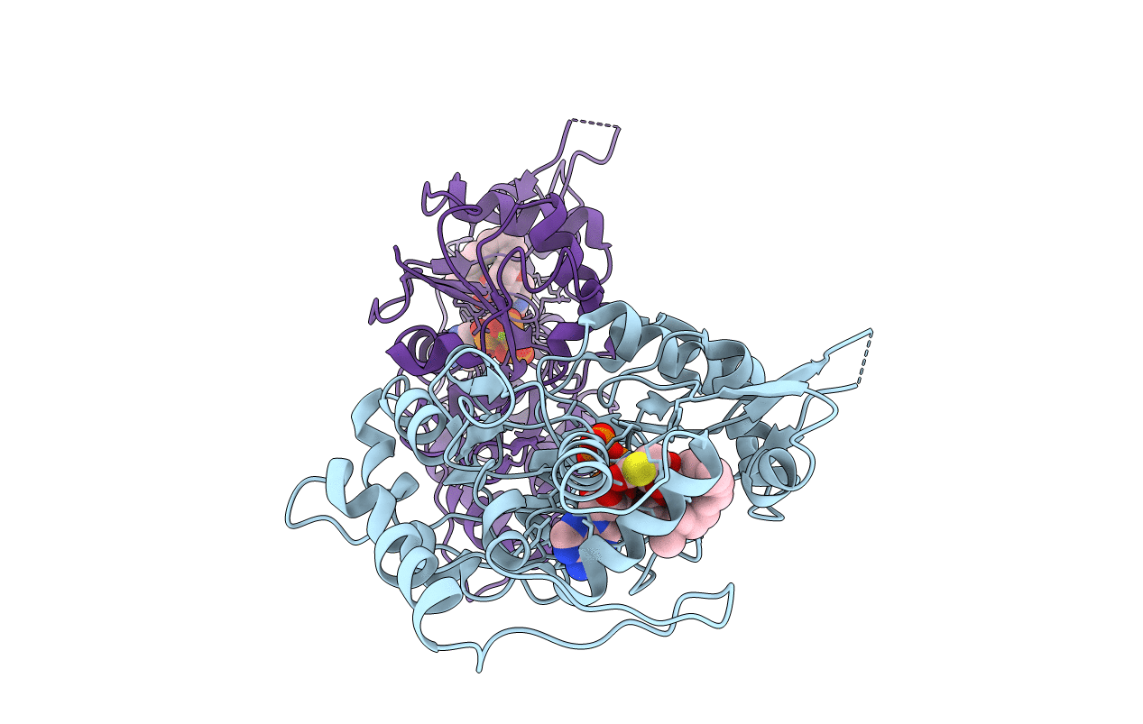
Title:
Actin Crystal Dynamics: Structural Implications for F-actin Nucleation, Polymerization and Branching Mediated by the Anti-parallel Dimer
Biological Source:
Source Organism(s):
Oryctolagus cuniculus (Taxon ID: 9986)
Method Details:
Experimental Method:
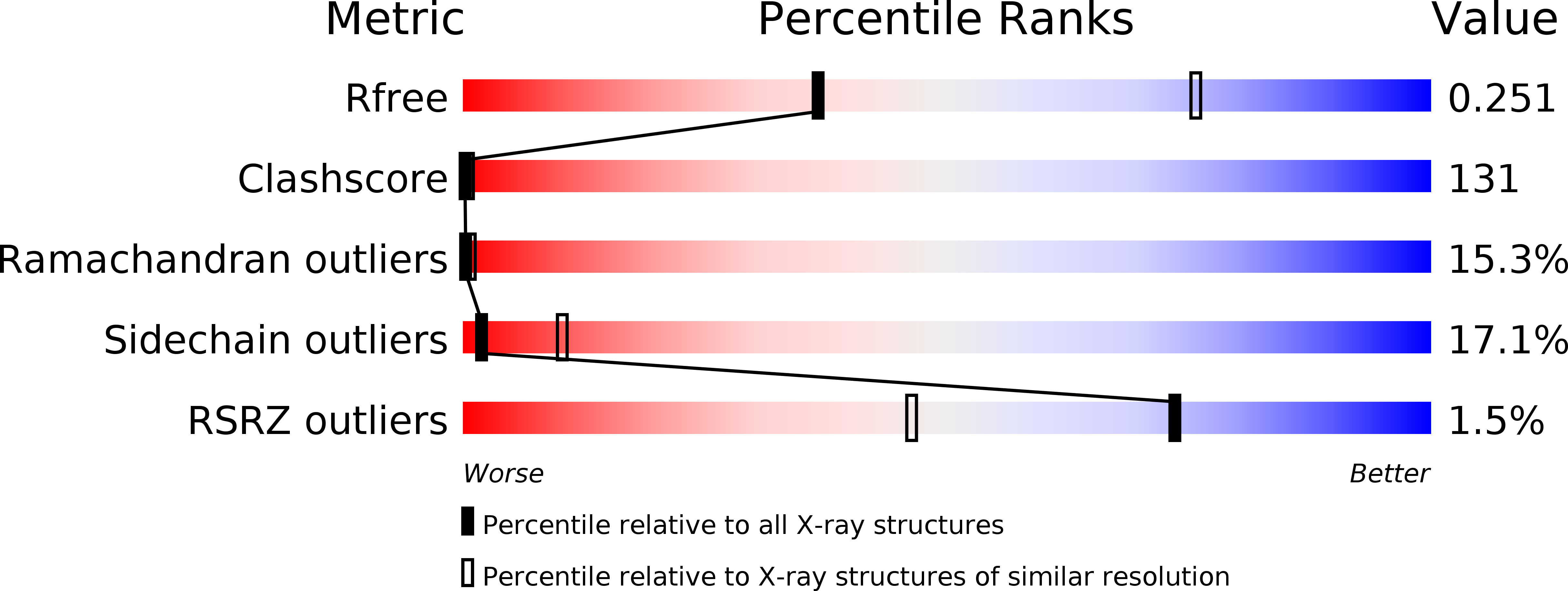
Resolution:
3.00 Å
R-Value Free:
0.26
R-Value Work:
0.19
Space Group:
P 43