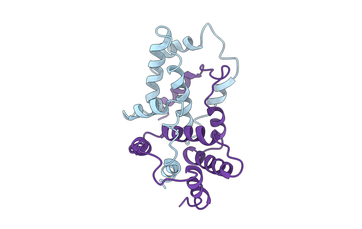
1RFB image
Deposition Date 1993-06-30
Release Date 1994-01-31
Last Version Date 2024-02-14
Entry Detail
PDB ID:
1RFB
Keywords:
Title:
CRYSTAL STRUCTURE OF RECOMBINANT BOVINE INTERFERON-GAMMA AT 3.0 ANGSTROMS RESOLUTION
Biological Source:
Source Organism:
Bos taurus (Taxon ID: 9913)
Method Details:
Experimental Method:
Resolution:
3.00 Å
R-Value Work:
0.19
R-Value Observed:
0.19
Space Group:
P 21 21 21
Macromolecular Entities
Structures with similar UniProt ID
Protein Blast
Polymer Type:polypeptide(L)
Molecule:INTERFERON-GAMMA
Gene (Uniprot):IFNG
Chain IDs:A, B
Chain Length:119
Number of Molecules:2
Biological Source:Bos taurus
Primary Citation
Structure of recombinant bovine interferon-gamma at 3.0 A resolution.
Acta Crystallogr.,Sect.D 49 513 521 (1993)
PMID: 15299487 DOI: 10.1107/S0907444993006924

Abstact

The three-dimensional crystal structure of recombinant bovine interferon-gamma was determined using the multiple isomorphous replacement method at 3.0 A and refined to an R factor of 19.2%. This protein crystallizes in space group P2(1)2(1)2(1) with unit-cell parameters of a = 42.8, b = 79.9 and c = 85.4 A. There is one functional dimer in the asymmetric unit. The two polypeptide chains are related by a non-crystallographic twofold symmetry axis. The secondary structure is predominantly alpha-helical with extensive interdigitation of the alpha-helical segments of the polypeptide chains that make up the dimer. The secondary structure, tertiary structure and topology of this molecule are identical to the previously reported structures of recombinant rabbit interferon-gamma and recombinant human interferon-gamma. The molecular topology is also similar to that of murine interferon-beta. These structural similarities strongly indicate the presence of a unique topological feature (fold) among gamma-interferons from different species, and also among the different classes of interferons.

Legend

Protein

Chemical

Disease

Primary Citation of related structures