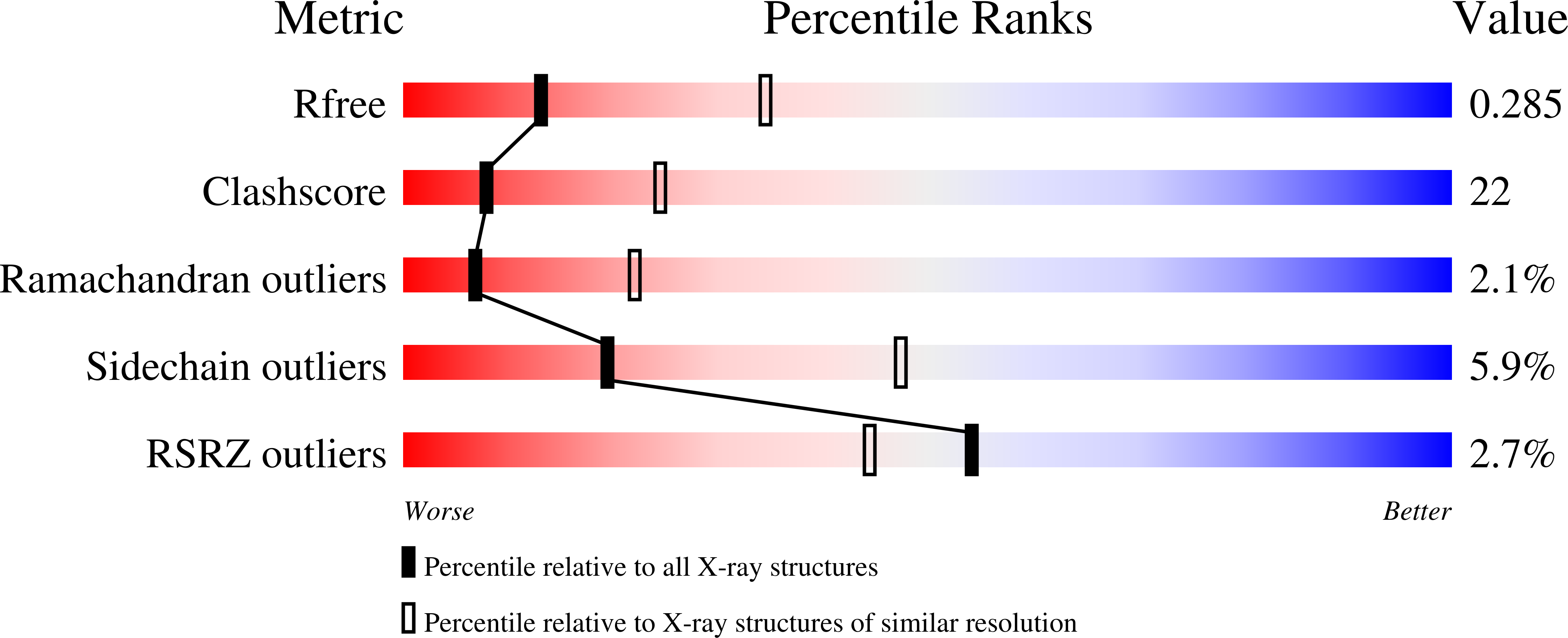
1RF0 image
Deposition Date 2003-11-07
Release Date 2004-03-16
Last Version Date 2024-11-13
Entry Detail
PDB ID:
1RF0
Keywords:
Title:
Crystal Structure of Fragment D of gammaE132A Fibrinogen
Biological Source:
Source Organism:
Homo sapiens (Taxon ID: 9606)
Host Organism:
Method Details:
Experimental Method:
Resolution:
2.81 Å
R-Value Free:
0.28
R-Value Work:
0.23
R-Value Observed:
0.23
Space Group:
P 21 21 21
Macromolecular Entities
Polymer Type:polypeptide(L)
Molecule:Fibrinogen alpha/alpha-E chain
Gene (Uniprot):FGA
Chain IDs:A, D
Chain Length:66
Number of Molecules:2
Biological Source:Homo sapiens
Polymer Type:polypeptide(L)
Molecule:Fibrinogen beta chain
Gene (Uniprot):FGB
Chain IDs:B, E
Chain Length:313
Number of Molecules:2
Biological Source:Homo sapiens
Polymer Type:polypeptide(L)
Molecule:Fibrinogen gamma chain
Gene (Uniprot):FGG
Mutations:E132A
Chain IDs:C, F
Chain Length:311
Number of Molecules:2
Biological Source:Homo sapiens
Modified Residue
Compound ID Chain ID Parent Comp ID Details 2D Image
ASN B ASN GLYCOSYLATION SITE
Primary Citation
Calcium-Binding Site beta2, Adjacent to the "b" Polymerization Site, Modulates Lateral Aggregation of Protofibrils during Fibrin Polymerization.
Biochemistry 43 2475 2483 (2004)
PMID: 14992585 DOI: 10.1021/bi0359978

Abstact

Structural analysis of recombinant fibrinogen fragment D revealed that the calcium-binding site (beta2-site) composed of residues BbetaAsp261, BbetaAsp398, BbetaGly263, and gammaGlu132 is modulated by the "B:b" interaction. To determine the beta2-site's role in polymerization, we engineered variant fibrinogen gammaE132A in which calcium binding to the beta2-site was disrupted by replacing glutamic acid at gamma132 with alanine. We compared polymerization of gammaE132A to normal fibrinogen as a function of calcium concentration. Polymerization of gammaE132A at concentrations of calcium <or=1 mM exhibited an uncharacteristic 2-3-fold increase in lateral aggregation and fiber thickness compared to normal fibrinogen, while polymerization of variant and normal were indistinguishable at 10 mM calcium. These results suggest that the beta2-site controls the extent of lateral aggregation. That is, when the calcium anchor (beta2-site) is eliminated before "B:b" interactions occur then lateral aggregation is enhanced. We solved structures of fragment D of gammaE132A fibrinogen (rfD-gammaE132A) with and without Gly-His-Arg-Pro-amide (GHRPam) and found no change to the global structure. X-ray diffraction data showed GHRPam binding in the "a" and "b" polymerization sites and that calcium could still bind to the beta2-site of gammaE132A fibrinogen at 70 mM calcium. We found that the gamma2 calcium-binding site (in loop gamma294-301) did not have calcium bound in the structure of fragment D of gammaE132A fibrinogen with GHRPam bound (rfD-gammaE132A+GH). Analysis of structures rfD-gammaE132A+GH and rfD-BbetaD398A+GH indicated that differences in calcium occupation of the gamma2-site resulted from minor conformational changes provoked by crystal packing and GHRPam binding to the "a" site did not directly modulate calcium binding to this site.

Legend

Protein

Chemical

Disease

Primary Citation of related structures