1RE3 image
Deposition Date 2003-11-06
Release Date 2004-05-25
Last Version Date 2024-10-30
Entry Detail
PDB ID:
1RE3
Keywords:
Title:
Crystal Structure of Fragment D of BbetaD398A Fibrinogen with the Peptide Ligand Gly-His-Arg-Pro-Amide
Biological Source:
Source Organism:
Homo sapiens (Taxon ID: 9606)
(Taxon ID: )
Host Organism:
Method Details:
Experimental Method:
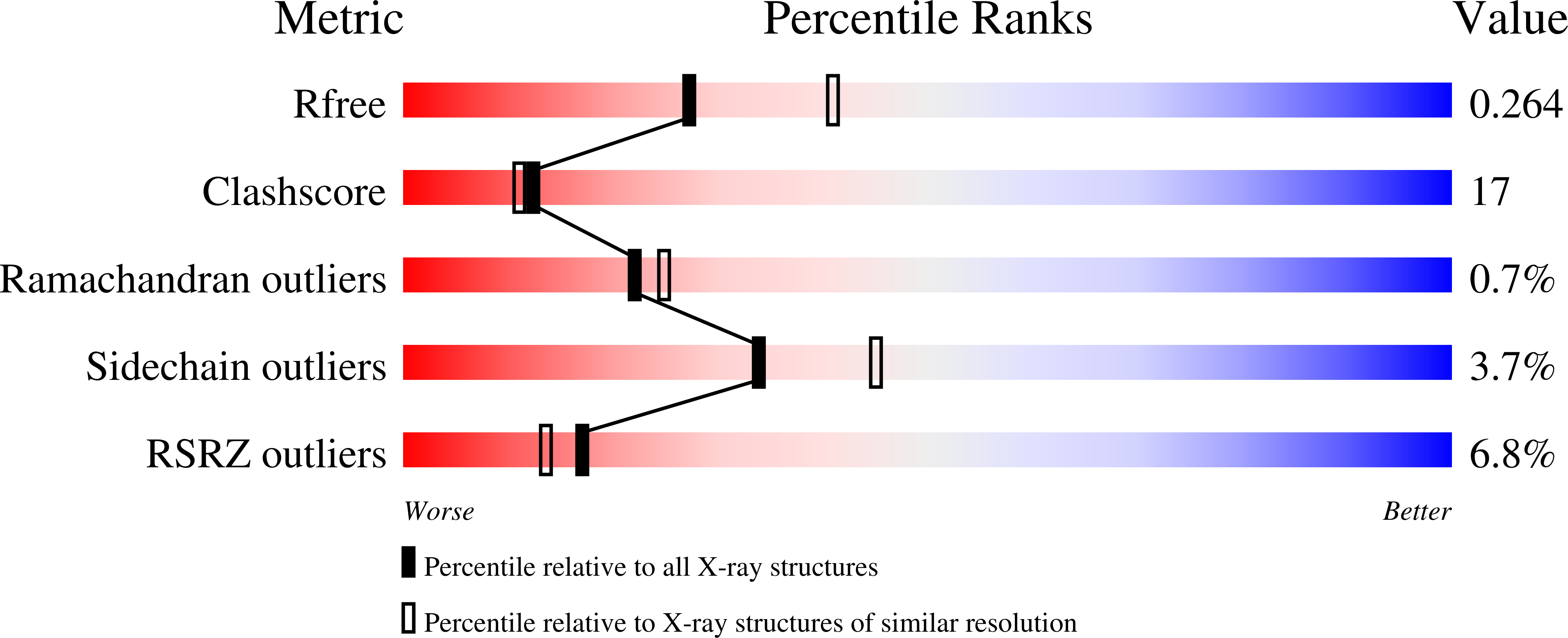
Resolution:
2.45 Å
R-Value Free:
0.26
R-Value Work:
0.22
R-Value Observed:
0.22
Space Group:
P 21 21 21
Macromolecular Entities
Polymer Type:polypeptide(L)
Molecule:Fibrinogen alpha/alpha-E chain
Gene (Uniprot):FGA
Chain IDs:A, D
Chain Length:66
Number of Molecules:2
Biological Source:Homo sapiens
Polymer Type:polypeptide(L)
Molecule:Fibrinogen beta chain
Gene (Uniprot):FGB
Mutations:D398A
Chain IDs:B, E
Chain Length:313
Number of Molecules:2
Biological Source:Homo sapiens
Polymer Type:polypeptide(L)
Molecule:Fibrinogen gamma chain
Gene (Uniprot):FGG
Chain IDs:C, F
Chain Length:311
Number of Molecules:2
Biological Source:Homo sapiens
Polymer Type:polypeptide(L)
Molecule:GHRP peptide
Chain IDs:G, H
Chain Length:4
Number of Molecules:2
Biological Source:
Modified Residue
Compound ID Chain ID Parent Comp ID Details 2D Image
ASN B ASN GLYCOSYLATION SITE
Ligand Molecules
Primary Citation
BbetaGlu397 and BbetaAsp398 but not BbetaAsp432 are required for "B:b" interactions.
Biochemistry 43 2465 2474 (2004)
PMID: 14992584 DOI: 10.1021/bi035996f

Abstact

We synthesized three fibrinogen variants, BbetaE397A, BbetaD398A, and BbetaD432A, with substitutions at positions identified in crystallographic studies as critical for binding the "B" peptide, Gly-His-Arg-Pro-amide (GHRPam), to the "b" polymerization site. We examined thrombin- and batroxobin-catalyzed polymerization by turbidity measurements and found that BbetaE397A and BbetaD398A were impaired while BbetaD432A was normal. Changes in polymerization as a function of calcium were similar for variant and normal fibrinogens. We determined crystal structures of fragment D from the variant BbetaD398A in the absence and presence of GHRPam. In the absence of peptide, the structure showed that the alanine substitution altered only specific local interactions, as alignment of the variant structure with the analogous normal structure resulted in an RMSD of 0.53 A over all atoms. The structure also showed reduced occupancy of the beta2 calcium-binding site that includes the side chain carbonyl of BbetaD398, suggesting that calcium was not bound at this site in our polymerization studies. In the presence of peptide, the structure showed that GHRPam was not bound in the "b" site and the conformational changes associated with peptide binding to normal fragment D did not occur. This structure also showed GHRPam bound in the "a" polymerization site, although in two different conformations. Calcium binding was associated with only one of these conformations, suggesting that calcium binding to the gamma2-site and an alternative peptide conformation were induced by crystal packing. We conclude that BbetaE397 and BbetaD398 are essential for the "B:b" interaction, while BbetaD432 is not.

Legend

Protein

Chemical

Disease

Primary Citation of related structures