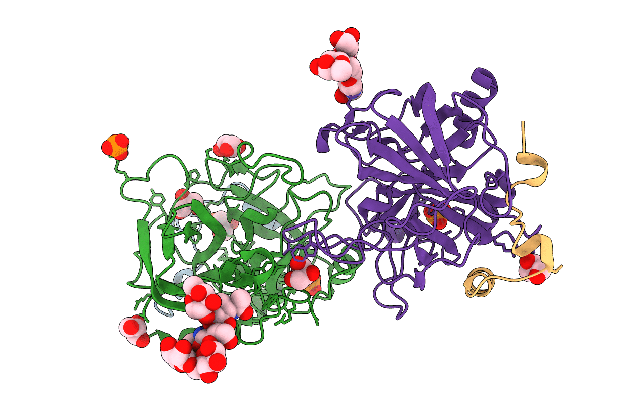
1RD3 image
Deposition Date 2003-11-05
Release Date 2004-05-04
Last Version Date 2024-11-13
Entry Detail
PDB ID:
1RD3
Keywords:
Title:
2.5A Structure of Anticoagulant Thrombin Variant E217K
Biological Source:
Source Organism:
Homo sapiens (Taxon ID: 9606)
Host Organism:
Method Details:
Experimental Method:
Resolution:
2.50 Å
R-Value Free:
0.25
R-Value Work:
0.22
Space Group:
P 21 21 21
Macromolecular Entities
Polymer Type:polypeptide(L)
Molecule:Prothrombin
Gene (Uniprot):F2
Chain IDs:A, C
Chain Length:36
Number of Molecules:2
Biological Source:Homo sapiens
Polymer Type:polypeptide(L)
Molecule:Prothrombin
Gene (Uniprot):F2
Mutations:E217K
Chain IDs:B, D
Chain Length:259
Number of Molecules:2
Biological Source:Homo sapiens
Modified Residue
Compound ID Chain ID Parent Comp ID Details 2D Image
ASN B ASN GLYCOSYLATION SITE
Primary Citation
Crystal Structure of Anticoagulant Thrombin Variant E217K Provides Insights into Thrombin Allostery
J.Biol.Chem. 279 26387 26394 (2004)
PMID: 15075325 DOI: 10.1074/jbc.M402364200

Abstact

Thrombin is the ultimate protease of the blood clotting cascade and plays a major role in its own regulation. The ability of thrombin to exhibit both pro- and anti-coagulant properties has spawned efforts to turn thrombin into an anticoagulant for therapeutic purposes. This quest culminated in the identification of the E217K variant through scanning and saturation mutagenesis. The antithrombotic properties of E217K thrombin are derived from its inability to convert fibrinogen to a fibrin clot while maintaining its thrombomodulin-dependent ability to activate the anticoagulant protein C pathway. Here we describe the 2.5-A crystal structure of human E217K thrombin, which displays a dramatic restructuring of the geometry of the active site. Of particular interest is the repositioning of Glu-192, which hydrogen bonds to the catalytic Ser-195 and which results in the complete occlusion of the active site and the destruction of the oxyanion hole. Substrate binding pockets are further blocked by residues previously implicated in thrombin allostery. We have concluded that the E217K mutation causes the allosteric inactivation of thrombin by destabilizing the Na(+) binding site and that the structure thus may represent the Na(+)-free, catalytically inert "slow" form.

Legend

Protein

Chemical

Disease

Primary Citation of related structures