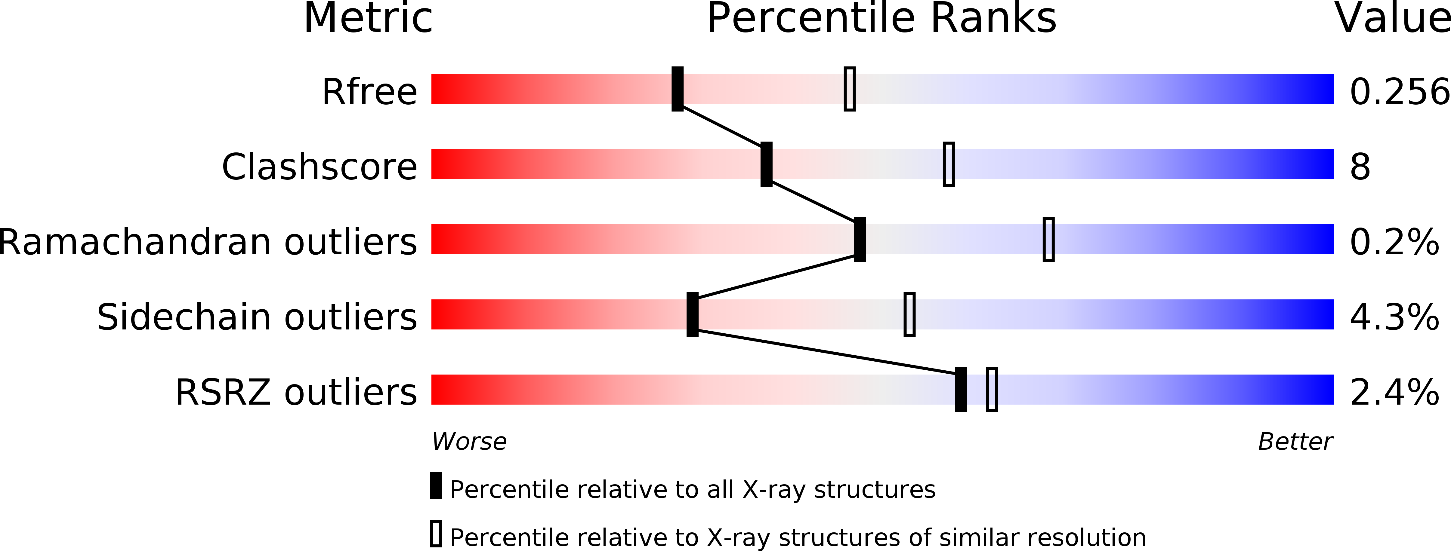
1RCW image
Deposition Date 2003-11-04
Release Date 2004-05-04
Last Version Date 2024-11-20
Entry Detail
PDB ID:
1RCW
Title:
Crystal structure of CT610 from Chlamydia trachomatis
Biological Source:
Source Organism:
Host Organism:
Method Details:
Experimental Method:
Resolution:
2.50 Å
R-Value Free:
0.25
R-Value Work:
0.19
R-Value Observed:
0.19
Space Group:
C 2 2 21
Macromolecular Entities
Structures with similar UniProt ID
Protein Blast
Polymer Type:polypeptide(L)
Molecule:CT610
Gene (Uniprot):CT_610
Chain IDs:A, B, C
Chain Length:231
Number of Molecules:3
Biological Source:Chlamydia trachomatis
Modified Residue
Compound ID Chain ID Parent Comp ID Details 2D Image
MSE A MET SELENOMETHIONINE
Ligand Molecules
Primary Citation
Structure of the Chlamydia protein CADD reveals a redox enzyme that modulates host cell apoptosis.
J.Biol.Chem. 279 29320 29324 (2004)
PMID: 15087448 DOI: 10.1074/jbc.M401268200

Abstact

The Chlamydia protein CADD (Chlamydia protein associating with death domains) has been implicated in the modulation of host cell apoptosis via binding to the death domains of tumor necrosis factor family receptors. Transfection of CADD into mammalian cells induces apoptosis. Here we present the CADD crystal structure, which reveals a dimer of seven-helix bundles. Each bundle contains a di-iron center adjacent to an internal cavity, forming an active site similar to that of methane mono-oxygenase hydrolase. We further show that CADD mutants lacking critical metal-coordinating residues are substantially less effective in inducing apoptosis but retain their ability to bind to death domains. We conclude that CADD is a novel redox protein toxin unique to Chlamydia species and propose that both its redox activity and death domain binding ability are required for its biological activity.

Legend

Protein

Chemical

Disease

Primary Citation of related structures