1RCO image
Deposition Date 1996-10-31
Release Date 1997-03-12
Last Version Date 2024-10-30
Entry Detail
PDB ID:
1RCO
Keywords:
Title:
SPINACH RUBISCO IN COMPLEX WITH THE INHIBITOR D-XYLULOSE-2,2-DIOL-1,5-BISPHOSPHATE
Biological Source:
Source Organism:
Method Details:
Experimental Method:
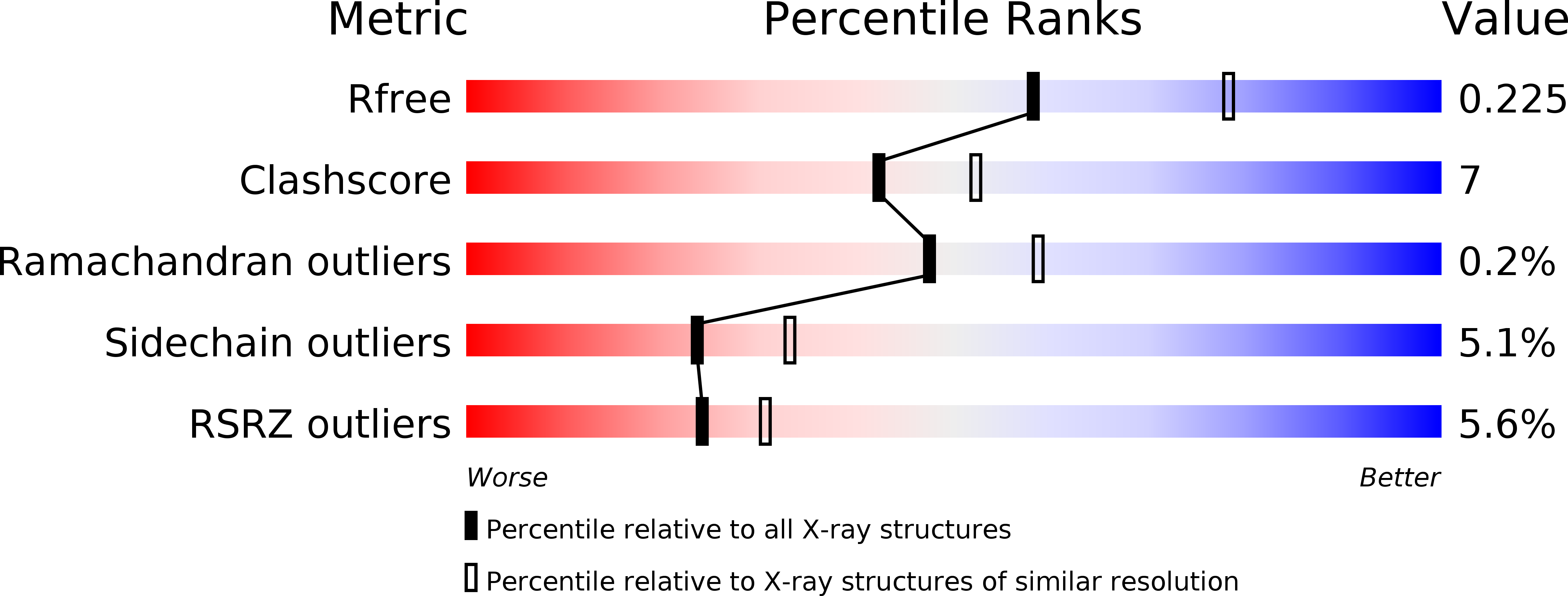
Resolution:
2.30 Å
R-Value Free:
0.24
R-Value Work:
0.23
R-Value Observed:
0.23
Space Group:
P 21 21 2
Macromolecular Entities
Polymer Type:polypeptide(L)
Molecule:RIBULOSE BISPHOSPHATE CARBOXYLASE/OXYGENASE
Gene (Uniprot):rbcL
Chain IDs:A (auth: L), C (auth: B), E, G (auth: H), I (auth: K), K (auth: O), M (auth: R), O (auth: V)
Chain Length:475
Number of Molecules:8
Biological Source:Spinacia oleracea
Polymer Type:polypeptide(L)
Molecule:RIBULOSE BISPHOSPHATE CARBOXYLASE/OXYGENASE
Gene (Uniprot):RBCS1
Chain IDs:B (auth: S), D (auth: C), F, H (auth: I), J (auth: M), L (auth: P), N (auth: T), P (auth: W)
Chain Length:123
Number of Molecules:8
Biological Source:Spinacia oleracea
Ligand Molecules
Primary Citation
A common structural basis for the inhibition of ribulose 1,5-bisphosphate carboxylase by 4-carboxyarabinitol 1,5-bisphosphate and xylulose 1,5-bisphosphate.
J.Biol.Chem. 271 32894 32899 (1996)
PMID: 8955130 DOI: 10.1074/jbc.271.51.32894

Abstact

Ribulose 1,5-bisphosphate carboxylase/oxygenase (Rubisco) catalyzes the carboxylation of ribulose 1,5-bisphosphate. The reaction catalyzed by Rubisco involves several steps, some of which can occur as partial reactions, forming intermediates that can be isolated. Analogues of these intermediates are potent inhibitors of the enzyme. We have studied the interactions with the enzyme of two inhibitors, xylulose 1,5-bisphosphate and 4-carboxyarabinitol 1,5-bisphosphate, by x-ray crystallography. Crystals of the complexes were formed by cocrystallization under activating conditions. In addition, 4-carboxyarabinitol 1,5-bisphosphate was soaked into preformed activated crystals of the enzyme. The result of these experiments was the release of the activating CO2 molecule as well as the metal ion from the active site when the inhibitors bound to the enzyme. Comparison with the structure of an activated complex of the enzyme indicates that the structural basis for the release of the activator groups is a distortion of the metal binding site due to the different geometry of the C-3 hydroxyl of the inhibitors. Both inhibitors induce closure of active site loops despite the inactivated state of the enzyme. Xylulose 1,5-bisphosphate binds in a hydrated form at the active site.

Legend

Protein

Chemical

Disease

Primary Citation of related structures