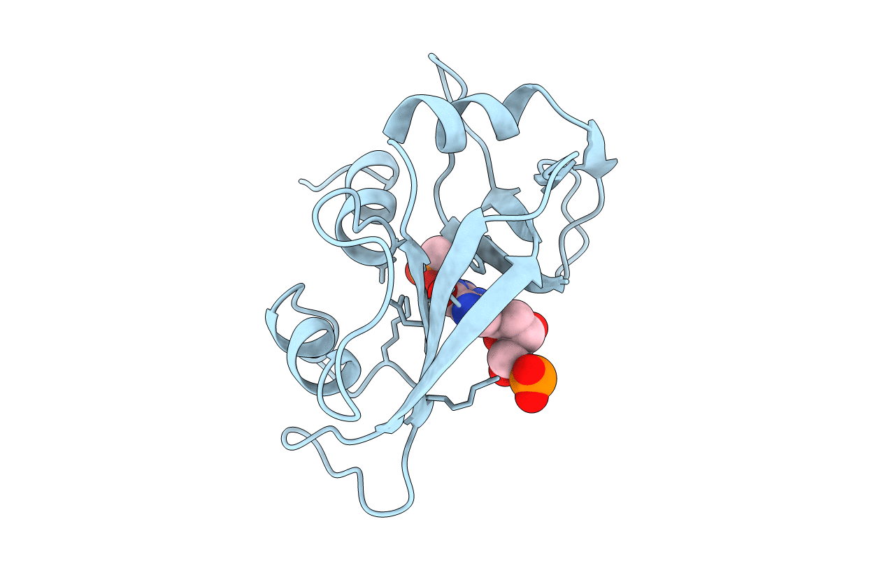
1RCA image
Entry Detail
PDB ID:
1RCA
Title:
STRUCTURE OF THE CRYSTALLINE COMPLEX OF DEOXYCYTIDYLYL-3',5'-GUANOSINE (3',5'-DCPDG) CO-CRYSTALISED WITH RIBONUCLEASE AT 1.9 ANGSTROMS RESOLUTION. RETROBINDING IN PANCREATIC RNASEA IS INDEPENDENT OF MODE OF INHIBITOR INTROMISSION
Biological Source:
Source Organism:
PDB Version:
Deposition Date:
1994-07-18
Release Date:
1995-09-15
Method Details:
Experimental Method:
Resolution:
1.90 Å
R-Value Observed:
0.21
Space Group:
P 1 21 1
Macromolecular Entities
Polymer Type:polypeptide(L)
Description:RIBONUCLEASE A
Chain IDs:A
Chain Length:124
Number of Molecules:1
Biological Source:Bos taurus
Primary Citation
Structure of the crystalline complex of deoxycytidylyl-3',5'-guanosine (3',5'-dCpdG) cocrystallized with ribonuclease at 1.9 A resolution.
Acta Crystallogr.,Sect.D 51 767 771 (1995)
PMID: 15299807 DOI: 10.1107/S0907444995001570

Abstact

The X-ray structure of bovine ribonuclease A cocrystallized with the dinucleotide deoxycytidylyl-3',5'-guanosine has been determined at 1.9 A resolution and refined by restrained least squares to R = 0.218 for 7807 reflections. The structure established that the recently observed retrobound mode of attachment of substrate analogues cytidylyl-2',5'-guanosine and deoxycytidylyl-3',5'-guanosine found in soaked RNase A crystals is also present in the cocrystallized complex. Retrobinding is thus unlikely to be the result of restrictions imposed by the crystalline environment as the ligands soak into the lattice but rather a phenomenon specific to small nucleotides containing guanine.

Legend

Protein

Chemical

Disease

Primary Citation of related structures