1RBL image
Deposition Date 1993-05-12
Release Date 1994-06-22
Last Version Date 2024-06-05
Entry Detail
PDB ID:
1RBL
Keywords:
Title:
STRUCTURE DETERMINATION AND REFINEMENT OF RIBULOSE 1,5 BISPHOSPHATE CARBOXYLASE(SLASH)OXYGENASE FROM SYNECHOCOCCUS PCC6301
Biological Source:
Source Organism:
Method Details:
Experimental Method:
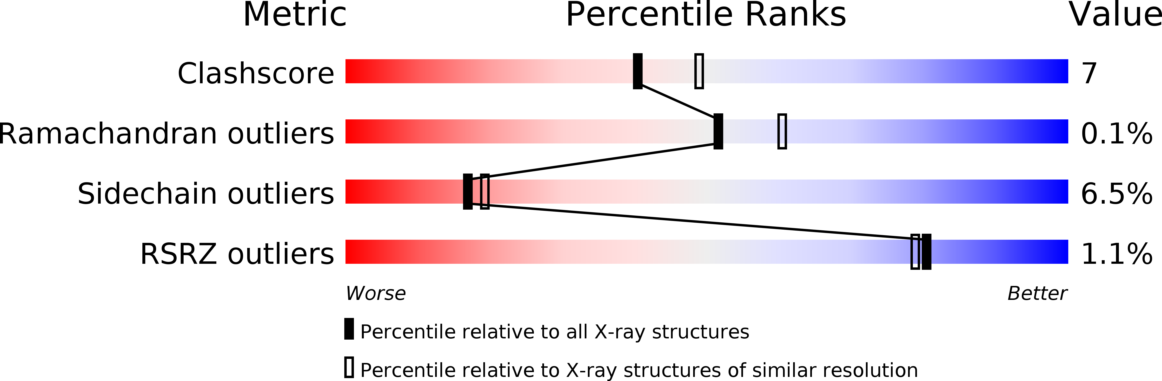
Resolution:
2.20 Å
R-Value Work:
0.2
R-Value Observed:
0.2
Space Group:
P 21 21 21
Macromolecular Entities
Structures with similar UniProt ID
Protein Blast
Polymer Type:polypeptide(L)
Molecule:RIBULOSE 1,5 BISPHOSPHATE CARBOXYLASE/OXYGENASE (LARGE CHAIN)
Gene (Uniprot):cbbL
Chain IDs:A, C (auth: B), E (auth: C), G (auth: D), I (auth: E), K (auth: F), M (auth: G), O (auth: H)
Chain Length:467
Number of Molecules:8
Biological Source:Synechococcus elongatus
Structures with similar UniProt ID
Protein Blast
Polymer Type:polypeptide(L)
Molecule:RIBULOSE 1,5 BISPHOSPHATE CARBOXYLASE/OXYGENASE (SMALL CHAIN)
Gene (Uniprot):cbbS
Chain IDs:B (auth: M), D (auth: I), F (auth: N), H (auth: J), J (auth: O), L (auth: K), N (auth: P), P (auth: L)
Chain Length:109
Number of Molecules:8
Biological Source:Synechococcus elongatus
Primary Citation
Structure determination and refinement of ribulose 1,5-bisphosphate carboxylase/oxygenase from Synechococcus PCC6301.
Acta Crystallogr.,Sect.D 49 548 560 (1993)
PMID: 15299492 DOI: 10.1107/S090744499300530X

Abstact

The structure of an activated quaternary complex of ribulose 1,5-bisphosphate carboxylase/oxygenase (rubisco) from Synechococcus PCC6301 has been solved by molecular replacement. The protein crystallizes in an orthorhombic P2(1)2(1)2(1) unit cell with a complete L(8)S(8) complex consisting of 4608 residues (37 680 non-hydrogen atoms) in the asymmetric unit. Data were collected both on film and image plate using synchrotron radiation; there were 218 276 unique reflections in the final 2.2 A data set. The eightfold non-crystallographic symmetry could be used both to improve map quality and to reduce the computing requirements of refinement. The coordinates were refined using strict non-crystallographic symmetry constraints. The stereochemistry of the final model is good, and the model has an R value of 20.0% for the reflections between 7 and 2.2 A.

Legend

Protein

Chemical

Disease

Primary Citation of related structures