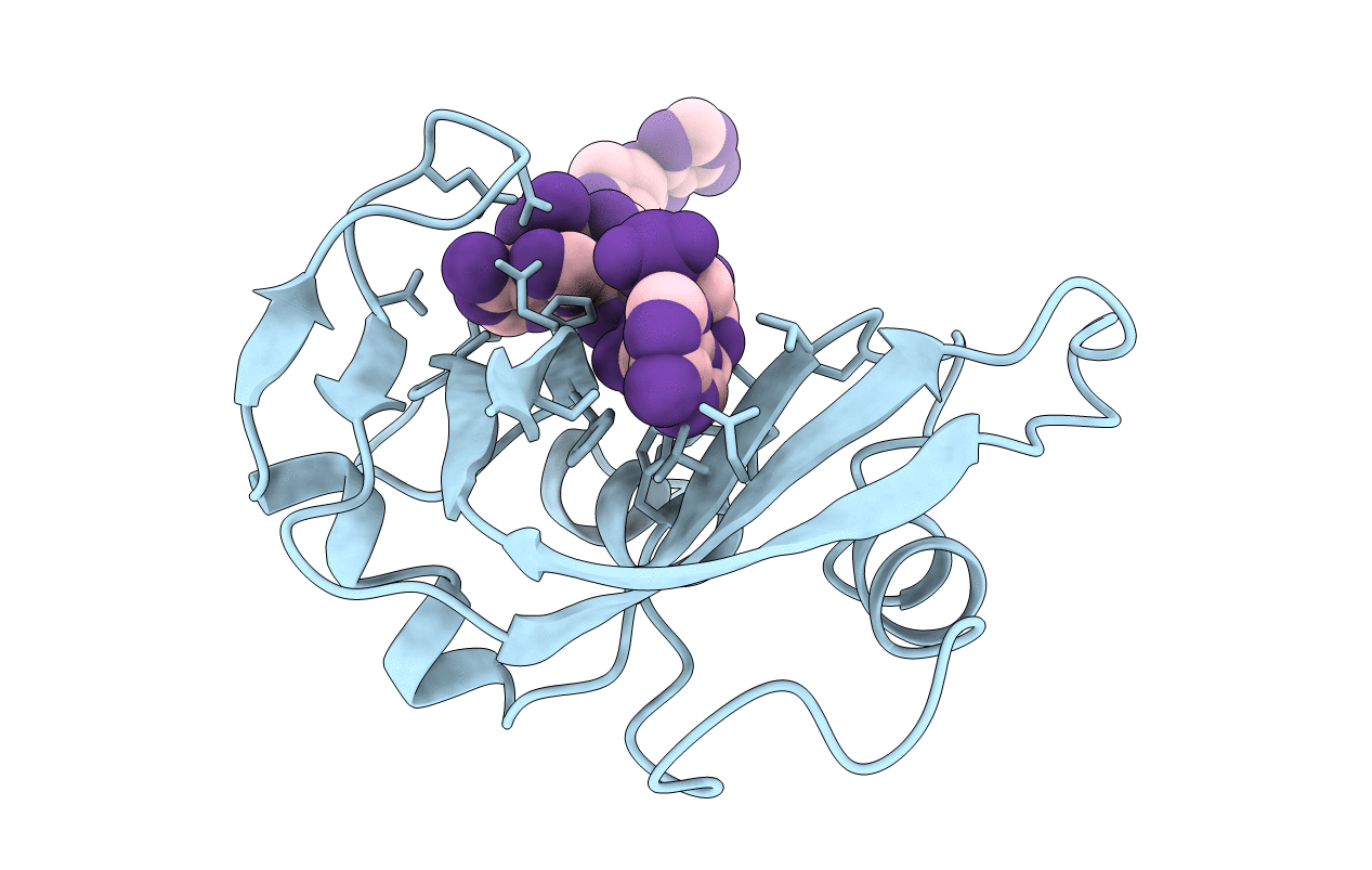
1RBJ image
Entry Detail
PDB ID:
1RBJ
Keywords:
Title:
RIBONUCLEASE B COMPLEX WITH D(TETRA-(DEOXY-ADENYLATE))
Biological Source:
Source Organism:
PDB Version:
Deposition Date:
1995-05-22
Release Date:
1995-12-07
Method Details:
Experimental Method:
Resolution:
2.70 Å
R-Value Observed:
0.16
Space Group:
P 41 21 2
Macromolecular Entities
Polymer Type:polypeptide(L)
Description:PROTEIN (RIBONUCLEASE B (E.C.3.1.27.5))
Chain IDs:B (auth: A)
Chain Length:124
Number of Molecules:1
Biological Source:Bos taurus
Ligand Molecules
Primary Citation
Structure of a ribonuclease B+d(pA)4 complex.
Acta Crystallogr.,Sect.D 52 160 164 (1996)
PMID: 15299737 DOI: 10.1107/S0907444995009127

Abstact

The structure of a tetragonal crystal of bovine pancreatic RNase B complexed with d(pA)(4) was determined by molecular replacement and difference Fourier methods. This crystal belongs to space group P4(1)2(1)2 and has unit-cell dimensions a = b = 44.5, c = 156.5 A. The model consists of the enzyme and a tetranucleotide with fractional occupancies, suggesting multiple modes of oligonucleotide binding. It does not include any polysaccharide residues or solvent molecules. After refinement at 2.7 A, the R value was 0.163 with acceptable stereochemistry. The model illustrates a set of well defined interactions for substrate binding, particularly between the central dinucleotide and the enzyme.

Legend

Protein

Chemical

Disease

Primary Citation of related structures