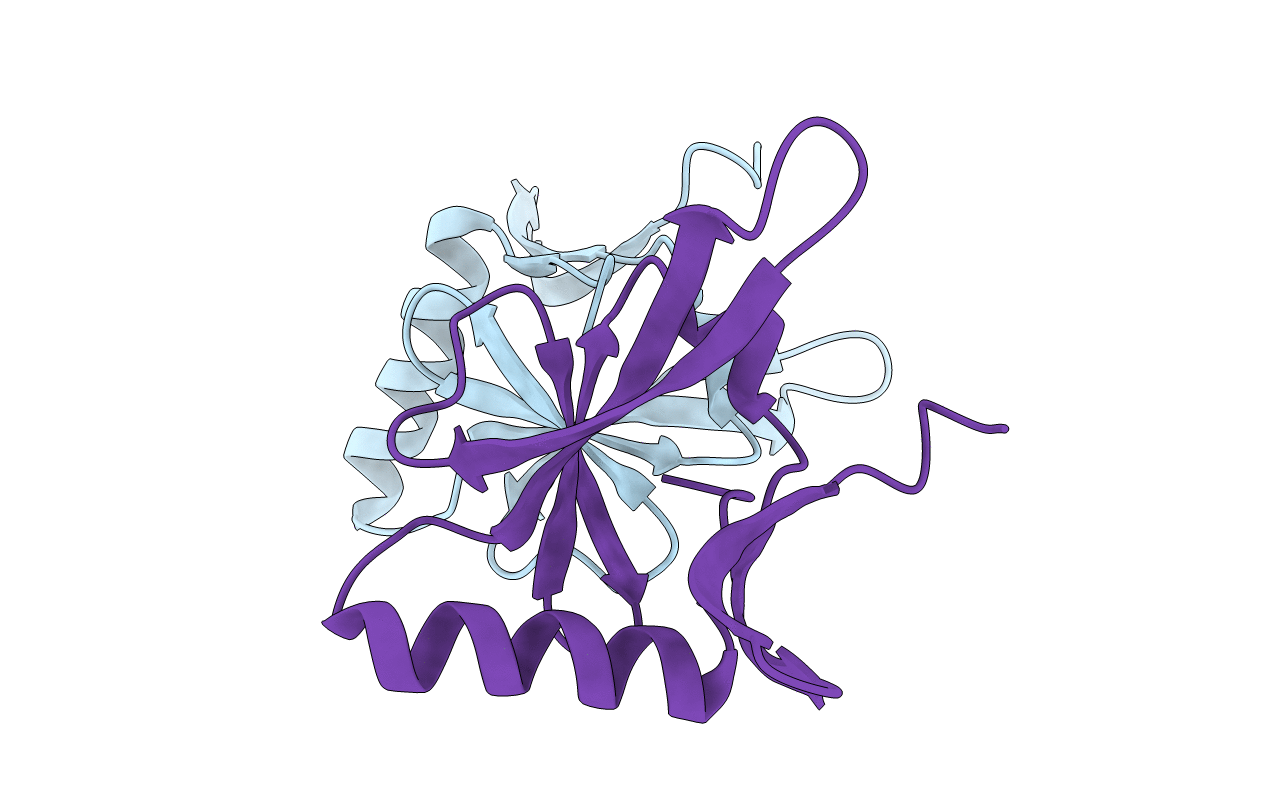
1R95 image
Deposition Date 2003-10-28
Release Date 2003-12-23
Last Version Date 2024-02-14
Entry Detail
PDB ID:
1R95
Keywords:
Title:
Crystal Structure of IscA (native)
Biological Source:
Source Organism:
Escherichia coli (Taxon ID: 562)
Host Organism:
Method Details:
Experimental Method:
Resolution:
2.65 Å
R-Value Free:
0.25
R-Value Work:
0.23
R-Value Observed:
0.23
Space Group:
P 62
Macromolecular Entities
Polymer Type:polypeptide(L)
Molecule:Protein yfhF
Gene (Uniprot):iscA
Chain IDs:A, B
Chain Length:118
Number of Molecules:2
Biological Source:Escherichia coli
Primary Citation
Crystal structure of the ancient, Fe-S scaffold IscA reveals a novel protein fold.
Biochemistry 43 133 139 (2004)
PMID: 14705938

Abstact

IscA belongs to an ancient family of proteins responsible for iron-sulfur cluster assembly in essential metabolic pathways preserved throughout evolution. We report here the 2.3 A resolution crystal structure of Escherichia coli IscA, a novel fold in which mixed beta-sheets form a compact alpha-beta sandwich domain. In contrast to the highly mobile secondary structural elements within the bacterial Fe-S scaffold protein IscU, a protein which is thought to have a similar function, the great majority of the amino acids that are conserved in IscA homologues are located in elements that constitute a well-ordered fold. However, the 10-residue C-terminal tail segment that contains two invariant cysteines critical for the Fe-S-binding function of a cyanobacterial (Synechocystis PCC) IscA homologue is not ordered in our structure. In addition, the crystal packing reveals a helical assembly that is constructed from two possible tetrameric oligomers of IscA.

Legend

Protein

Chemical

Disease

Primary Citation of related structures