1R85 image
Deposition Date 2003-10-23
Release Date 2004-07-20
Last Version Date 2024-04-03
Entry Detail
PDB ID:
1R85
Keywords:
Title:
Crystal structure of the extracellular xylanase from Geobacillus stearothermophilus T-6 (XT6): The WT enzyme (monoclinic form) at 1.45A resolution
Biological Source:
Source Organism:
Host Organism:
Method Details:
Experimental Method:
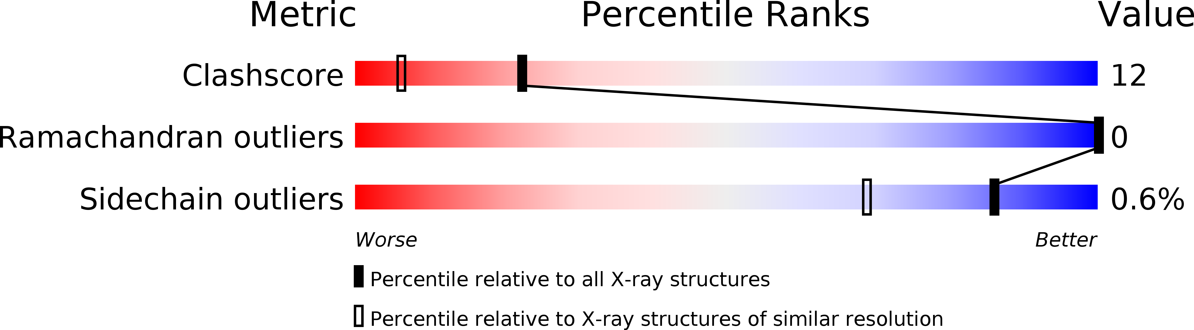
Resolution:
1.45 Å
R-Value Free:
0.18
R-Value Observed:
0.12
Space Group:
C 1 2 1
Macromolecular Entities
Structures with similar UniProt ID
Protein Blast
Polymer Type:polypeptide(L)
Molecule:Endo-1,4-beta-xylanase
Chain IDs:A
Chain Length:379
Number of Molecules:1
Biological Source:Geobacillus stearothermophilus
Primary Citation
Mapping glycoside hydrolase substrate subsites by isothermal titration calorimetry.
Proc.Natl.Acad.Sci.Usa 101 11275 11280 (2004)
PMID: 15277671 DOI: 10.1073/pnas.0404311101

Abstact

Relating thermodynamic parameters to structural and biochemical data allows a better understanding of substrate binding and its contribution to catalysis. The analysis of the binding of carbohydrates to proteins or enzymes is a special challenge because of the multiple interactions and forces involved. Isothermal titration calorimetry (ITC) provides a direct measure of binding enthalpy (DeltaHa) and allows the determination of the binding constant (free energy), entropy, and stoichiometry. In this study, we used ITC to elucidate the binding thermodynamics of xylosaccharides for two xylanases of family 10 isolated from Geobacillus stearothermophilus T-6. The change in the heat capacity of binding (DeltaCp = DeltaH/DeltaT) for xylosaccharides differing in one sugar unit was determined by using ITC measurements at different temperatures. Because hydrophobic stacking interactions are associated with negative DeltaCp, the data allow us to predict the substrate binding preference in the binding subsites based on the crystal structure of the enzyme. The proposed positional binding preference was consistent with mutants lacking aromatic binding residues at different subsites and was also supported by tryptophan fluorescence analysis.

Legend

Protein

Chemical

Disease

Primary Citation of related structures