Deposition Date
2003-10-23
Release Date
2003-11-11
Last Version Date
2024-10-30
Entry Detail
PDB ID:
1R84
Keywords:
Title:
NMR structure of the 13-cis-15-syn retinal in dark_adapted bacteriorhodopsin
Biological Source:
Source Organism(s):
Halobacterium salinarum (Taxon ID: 2242)
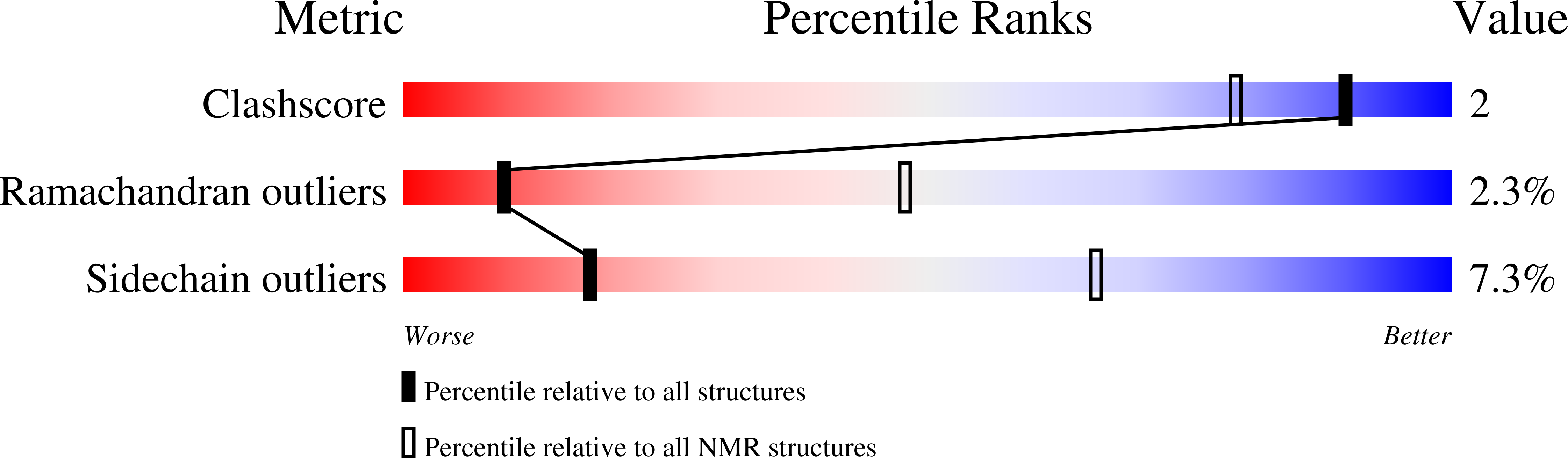
Method Details:
Experimental Method:
Conformers Calculated:
100
Conformers Submitted:
12
Selection Criteria:
rmsd between observed and calulated chemical shifts