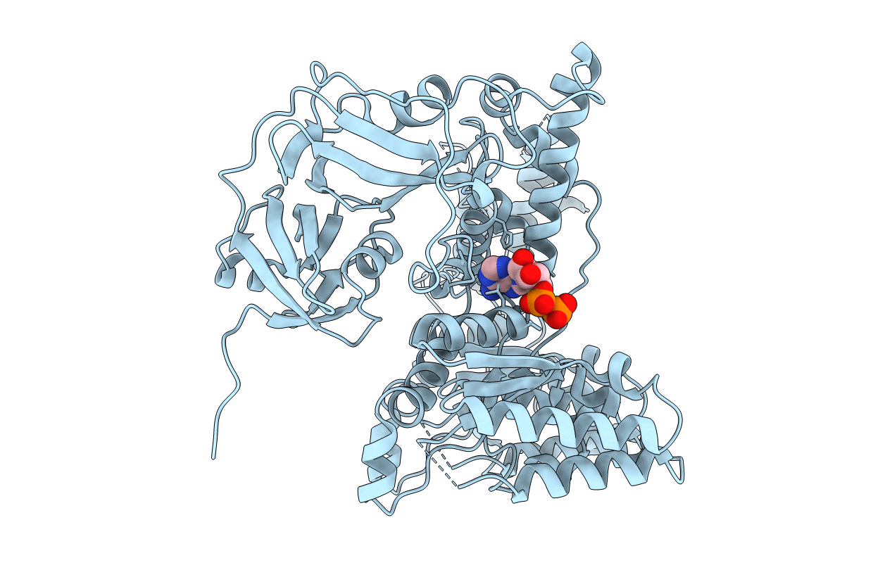
1R7R image
Deposition Date 2003-10-22
Release Date 2003-12-16
Last Version Date 2023-08-23
Entry Detail
PDB ID:
1R7R
Title:
The crystal structure of murine p97/VCP at 3.6A
Biological Source:
Source Organism:
Mus musculus (Taxon ID: 10090)
Host Organism:
Method Details:
Experimental Method:
Resolution:
3.60 Å
R-Value Free:
0.35
R-Value Work:
0.32
R-Value Observed:
0.32
Space Group:
P 6 2 2
Macromolecular Entities
Structures with similar UniProt ID
Protein Blast
Polymer Type:polypeptide(L)
Molecule:Transitional endoplasmic reticulum ATPase
Gene (Uniprot):Vcp
Chain IDs:A
Chain Length:816
Number of Molecules:1
Biological Source:Mus musculus
Ligand Molecules
Primary Citation
The crystal structure of murine p97/VCP at 3.6A
J.Struct.Biol. 144 337 348 (2003)
PMID: 14643202 DOI: 10.1016/j.jsb.2003.10.007

Abstact

p97/VCP is a member of the AAA ATPase family and has roles in both membrane fusion and ubiquitin dependent protein degradation. Here, we present a 3.6A crystal structure of murine p97 in which D2 domain has been modelled as poly-alanine and the remaining approximately 100 residues are absent. The resulting structure illustrates a head-to-tail packing arrangement of the two p97 AAA domains in a natural hexameric state with D1 ADP bound and D2 nucleotide free. The head-to-tail packing arrangement observed in this structure is in contrast to our previously predicted tail-to-tail packing model. The linker between the D1 and D2 domains is partially disordered, suggesting a flexible nature. Normal mode analysis of the crystal structure suggests anti-correlated motions and distinct conformational states of the two AAA domains.

Legend

Protein

Chemical

Disease

Primary Citation of related structures