1R7H image
Deposition Date 2003-10-21
Release Date 2004-05-04
Last Version Date 2024-10-30
Entry Detail
PDB ID:
1R7H
Title:
NrdH-redoxin of Corynebacterium ammoniagenes forms a domain-swapped dimer
Biological Source:
Source Organism:
Host Organism:
Method Details:
Experimental Method:
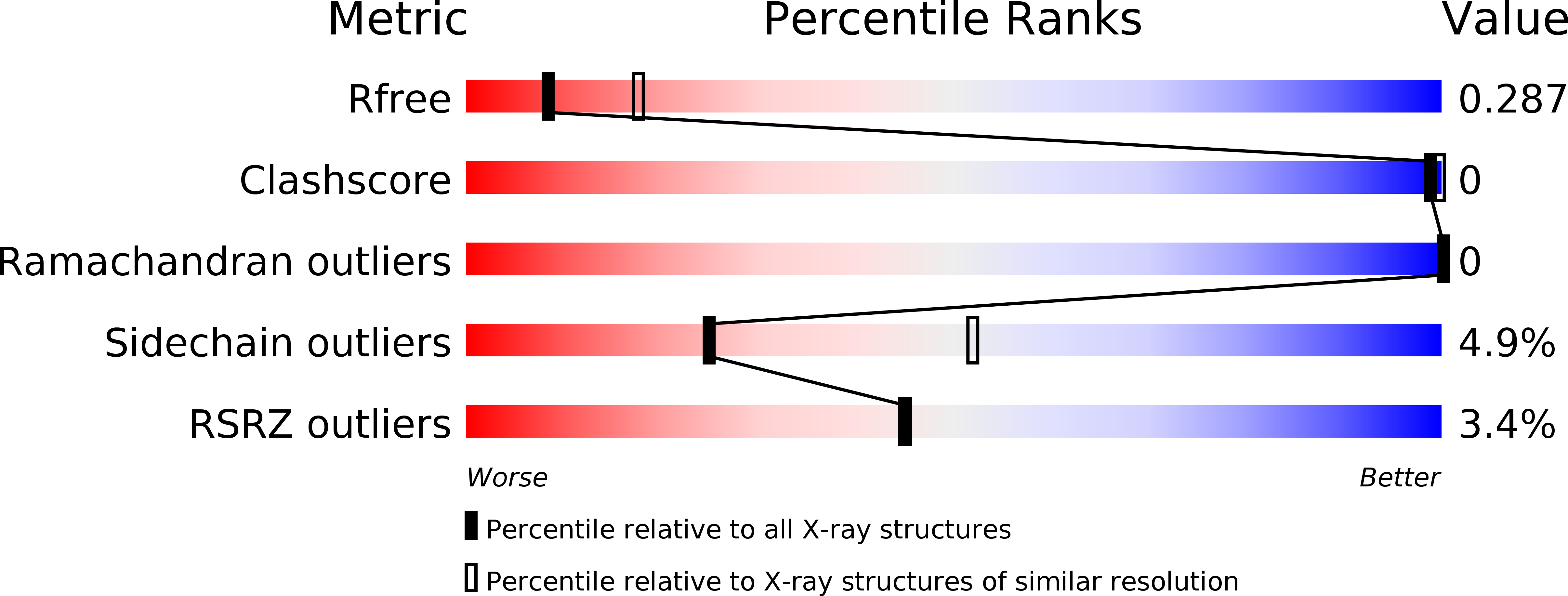
Resolution:
2.69 Å
R-Value Free:
0.28
R-Value Work:
0.24
R-Value Observed:
0.24
Space Group:
P 63
Macromolecular Entities
Structures with similar UniProt ID
Protein Blast
Polymer Type:polypeptide(L)
Molecule:NrdH-redoxin
Gene (Uniprot):nrdH
Chain IDs:A, B
Chain Length:75
Number of Molecules:2
Biological Source:Corynebacterium ammoniagenes
Primary Citation
NrdH-redoxin of Corynebacterium ammoniagenes forms a domain-swapped dimer.
Proteins 55 613 619 (2004)
PMID: 15103625 DOI: 10.1002/prot.20126

Abstact

NrdH-redoxins constitute a family of small redox proteins, which contain a conserved CXXC sequence motif, and are characterized by a glutaredoxin-like amino acid sequence but a thioredoxin-like activity profile. Here we report the structure of Corynebacterium ammoniagenes NrdH at 2.7 A resolution, determined by molecular replacement using E. coli NrdH as model. The structure is the first example of a domain-swapped dimer from the thioredoxin family. The domain-swapped structure is formed by an inter-chain two-stranded anti-parallel beta-sheet and is stabilized by electrostatic interactions at the dimer interface. Size exclusion chromatography, and MALDI-ESI experiments revealed however, that the protein exists as a monomer in solution. Similar to E. coli NrdH-redoxin and thioredoxin, C. ammoniagenes NrdH-redoxin has a wide hydrophobic pocket at the surface that could be involved in binding to thioredoxin reductase. However, the loop between alpha2 and beta3, which is complementary to a crevice in the reductase in the thioredoxin-thioredoxin reductase complex, is the hinge for formation of the swapped dimer in C. ammoniagenes NrdH-redoxin. C. ammoniagenes NrdH-redoxin has the highly conserved sequence motif W61-S-G-F-R-P-[DE]67 which is unique to the NrdH-redoxins and which determines the orientation of helix alpha3. An extended hydrogen-bond network, similar to that in E. coli NrdH-redoxin, determines the conformation of the loop formed by the conserved motif.

Legend

Protein

Chemical

Disease

Primary Citation of related structures