1R64 image
Deposition Date 2003-10-14
Release Date 2004-03-09
Last Version Date 2024-10-16
Entry Detail
PDB ID:
1R64
Title:
The 2.2 A crystal structure of Kex2 protease in complex with Ac-Arg-Glu-Lys-boroArg peptidyl boronic acid inhibitor
Biological Source:
Source Organism:
Method Details:
Experimental Method:
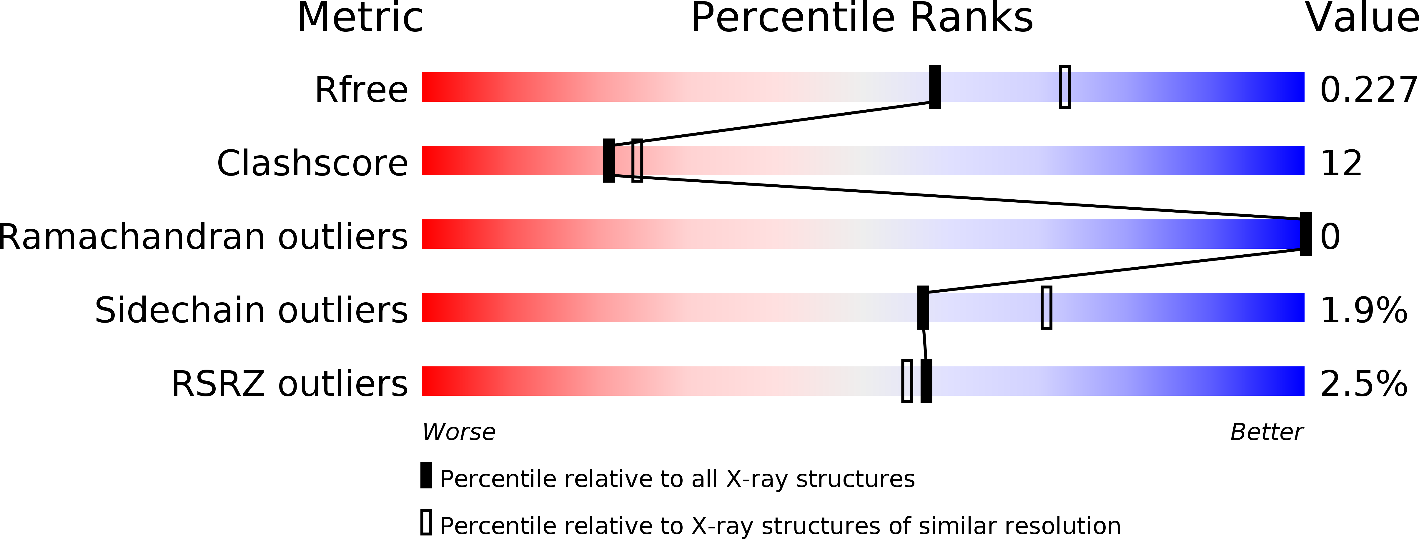
Resolution:
2.20 Å
R-Value Free:
0.23
R-Value Work:
0.19
R-Value Observed:
0.19
Space Group:
P 65 2 2
Macromolecular Entities
Structures with similar UniProt ID
Protein Blast
Polymer Type:polypeptide(L)
Molecule:Kexin
Gene (Uniprot):KEX2
Chain IDs:A, B
Chain Length:481
Number of Molecules:2
Biological Source:Saccharomyces cerevisiae
Polymer Type:polypeptide(L)
Molecule:Ac-Arg-Glu-Lys-boroArg peptide inhibitor
Chain IDs:C, D
Chain Length:5
Number of Molecules:2
Biological Source:
Modified Residue
Compound ID Chain ID Parent Comp ID Details 2D Image
ASN A ASN GLYCOSYLATION SITE
BOR C ARG ?
Peptide-like Molecules
PRD_000420
Primary Citation
Structural Basis for Differences in Substrate Selectivity in Kex2 and Furin Protein Convertases
Biochemistry 43 2412 2421 (2004)
PMID: 14992578 DOI: 10.1021/bi035849h

Abstact

Kex2 is the yeast prototype of a large family of serine proteases that are highly specific for cleavage of their peptide substrates C-terminal to paired basic sites. This paper reports the 2.2 A resolution crystal structure of ssKex2 in complex with an Ac-Arg-Glu-Lys-Arg peptidyl boronic acid inhibitor (R = 19.7, R(free) = 23.4). By comparison of this structure with the structure of the mammalian homologue furin [Henrich, S., et al. (2003) Nat. Struct. Biol. 10, 520-526], we suggest a structural basis for the differences in substrate recognition at the P(2) and P(4) positions between Kex2 and furin and provide a structural rationale for the lack of P(6) recognition in Kex2. In addition, several monovalent cation binding sites are identified, and a mechanism of activation of Kex2 by potassium ion is proposed.

Legend

Protein

Chemical

Disease

Primary Citation of related structures