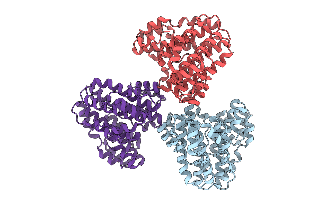
1R5Z image
Deposition Date 2003-10-14
Release Date 2004-01-13
Last Version Date 2024-11-13
Entry Detail
PDB ID:
1R5Z
Keywords:
Title:
Crystal Structure of Subunit C of V-ATPase
Biological Source:
Source Organism:
Host Organism:
Method Details:
Experimental Method:
Resolution:
1.95 Å
R-Value Free:
0.24
R-Value Work:
0.19
R-Value Observed:
0.19
Space Group:
P 61
Macromolecular Entities
Structures with similar UniProt ID
Protein Blast
Polymer Type:polypeptide(L)
Molecule:V-type ATP synthase subunit C
Gene (Uniprot):atpC
Chain IDs:A, B, C
Chain Length:323
Number of Molecules:3
Biological Source:Thermus thermophilus
Primary Citation
Crystal structure of a central stalk subunit C and reversible association/dissociation of vacuole-type ATPase.
Proc.Natl.Acad.Sci.Usa 101 59 64 (2004)
PMID: 14684831 DOI: 10.1073/pnas.0305165101

Abstact

The vacuole-type ATPases (V-ATPases) exist in various intracellular compartments of eukaryotic cells to regulate physiological processes by controlling the acidic environment. The crystal structure of the subunit C of Thermus thermophilus V-ATPase, homologous to eukaryotic subunit d of V-ATPases, has been determined at 1.95-A resolution and located into the holoenzyme complex structure obtained by single particle analysis as suggested by the results of subunit cross-linking experiments. The result shows that V-ATPase is substantially longer than the related F-type ATPase, due to the insertion of subunit C between the V(1) (soluble) and the V(o) (membrane bound) domains. Subunit C, attached to the V(o) domain, seems to have a socket like function in attaching the central-stalk subunits of the V(1) domain. This architecture seems essential for the reversible association/dissociation of the V(1) and the V(o) domains, unique for V-ATPase activity regulation.

Legend

Protein

Chemical

Disease

Primary Citation of related structures