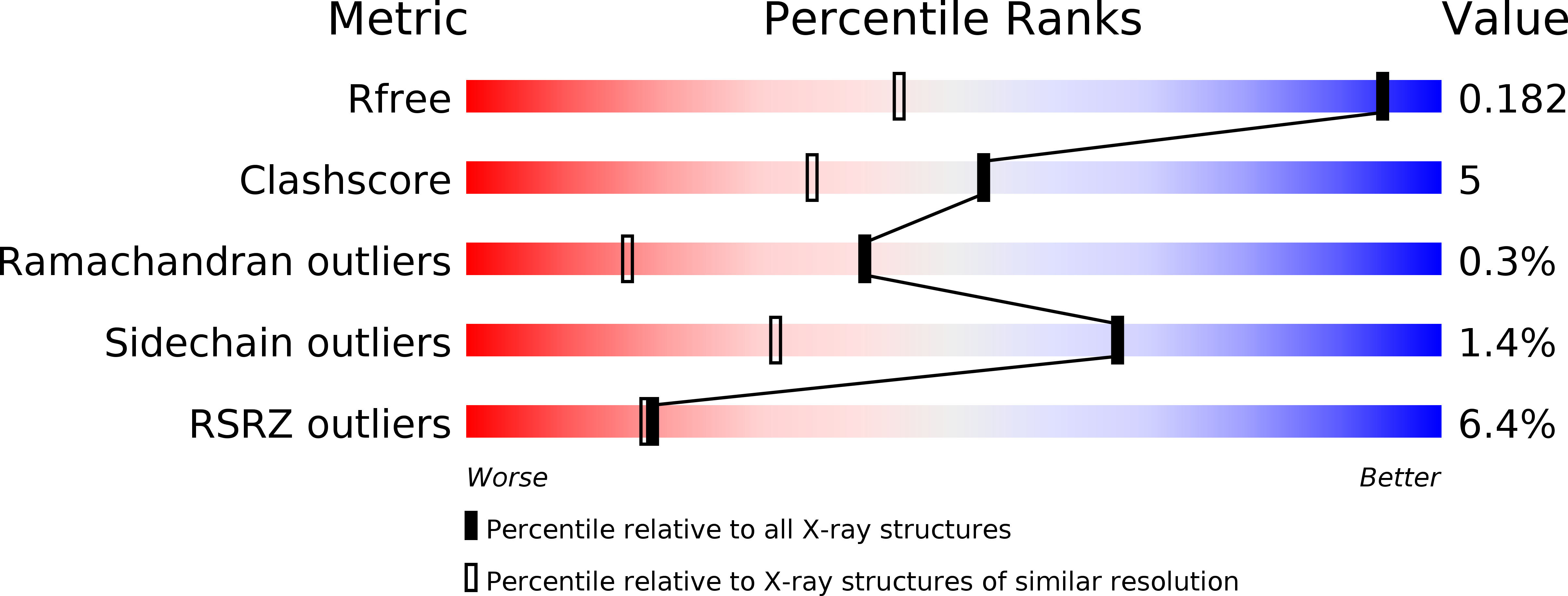
1R5Y image
Deposition Date 2003-10-13
Release Date 2004-04-13
Last Version Date 2023-11-08
Entry Detail
PDB ID:
1R5Y
Keywords:
Title:
Crystal Structure of TGT in complex with 2,6-Diamino-3H-Quinazolin-4-one Crystallized at PH 5.5
Biological Source:
Source Organism:
Method Details:
Experimental Method:
Resolution:
1.20 Å
R-Value Free:
0.19
R-Value Work:
0.17
R-Value Observed:
0.17
Space Group:
C 1 2 1
Macromolecular Entities
Structures with similar UniProt ID
Protein Blast
Polymer Type:polypeptide(L)
Molecule:Queuine tRNA-ribosyltransferase
Gene (Uniprot):tgt
Chain IDs:A
Chain Length:386
Number of Molecules:1
Biological Source:Zymomonas mobilis
Primary Citation
Crystallographic Study of Inhibitors of tRNA-guanine Transglycosylase Suggests a New Structure-based Pharmacophore for Virtual Screening.
J.Mol.Biol. 338 55 75 (2004)
PMID: 15050823 DOI: 10.1016/j.jmb.2004.02.019

Abstact

The enzyme tRNA-guanine transglycosylase (TGT) is involved in the pathogenicity of Shigellae. As the crystal structure of this protein is known, it is a putative target for the structure-based design of inhibitors. Here we report a crystallographic study of several new ligands exhibiting a 2,6-diamino-3H-quinazolin-4-one scaffold, which has been shown recently to be a promising template for TGT-inhibitors. Crystal structure analysis of these complexes has revealed an unexpected movement of the side-chain of Asp102. A detailed analysis of the water network disrupted by this rotation has lead to the derivation of a new composite pharmacophore. A virtual screening has been performed based on this pharmacophore hypothesis and several new inhibitors of micromolar binding affinity with new skeletons have been discovered.

Legend

Protein

Chemical

Disease

Primary Citation of related structures