Deposition Date
2003-10-10
Release Date
2003-11-25
Last Version Date
2024-11-20
Entry Detail
PDB ID:
1R5L
Keywords:
Title:
Crystal Structure of Human Alpha-Tocopherol Transfer Protein Bound to its Ligand
Biological Source:
Source Organism(s):
Homo sapiens (Taxon ID: 9606)
Expression System(s):
Method Details:
Experimental Method:
Resolution:
1.50 Å
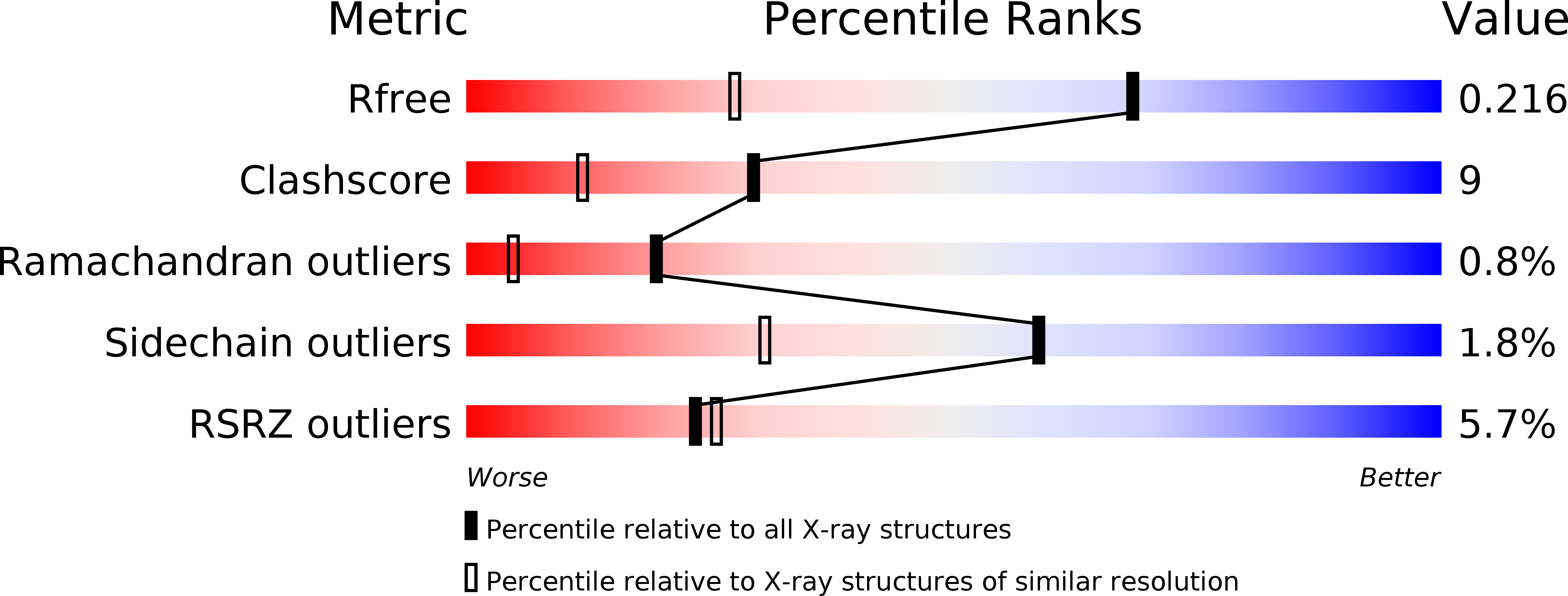
R-Value Free:
0.21
R-Value Work:
0.18
R-Value Observed:
0.18
Space Group:
P 21 21 21