1R48 image
Deposition Date 2003-10-03
Release Date 2003-12-23
Last Version Date 2024-05-22
Entry Detail
PDB ID:
1R48
Title:
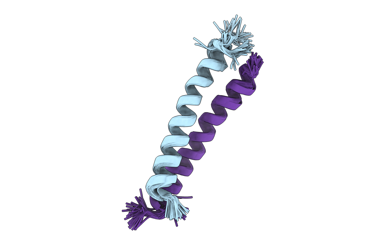
Solution structure of the C-terminal cytoplasmic domain residues 468-497 of Escherichia coli protein ProP
Biological Source:
Source Organism:
Method Details:
Experimental Method:
Conformers Calculated:
63
Conformers Submitted:
51
Selection Criteria:
structures with favorable non-bond energy
Macromolecular Entities
Structures with similar UniProt ID
Protein Blast
Polymer Type:polypeptide(L)
Molecule:Proline/betaine transporter
Chain IDs:A, B
Chain Length:33
Number of Molecules:2
Biological Source:
Ligand Molecules
Primary Citation
Solution Structure of the C-terminal Antiparallel Coiled-coil Domain from Escherichia coli Osmosensor ProP.
J.Mol.Biol. 334 1063 1076 (2003)
PMID: 14643666 DOI: 10.1016/j.jmb.2003.10.020

Abstact

Bacteria respond to increasing medium osmolality by accumulating organic solutes that are compatible with cellular functions. Transporter ProP of Escherichia coli, a proton symporter and a member of the major facilitator superfamily, senses osmotic shifts and responds by importing osmolytes such as glycine betaine. ProP contains a cytoplasmic, C-terminal extension that is essential for its activity. A peptide corresponding to the C-terminal extension of ProP forms a homodimeric alpha-helical coiled-coil even though some of its heptad a positions are not occupied by hydrophobic amino acid residues. Unexpectedly, amino acid replacement R488I, occurring at a heptad a position, destabilized the coiled-coil formed by the ProP peptide and attenuated the response of the intact transporter to osmotic upshifts in vivo. Thus, ProP was proposed to dimerize via an antiparallel coiled-coil. We used nuclear magnetic resonance (NMR) spectroscopy to determine the structure of the synthetic peptide corresponding to residues 468-497 of ProP. This region did form an antiparallel coil-coil in which critical residue R488 specifies the antiparallel coiled-coil orientation by forming stabilizing salt-bridges. Charged residues (both acidic and basic) are clustered on the c/g surface of the coiled-coil whereas polar residues are distributed on the b/e surface. This causes the structure to be bent, in contrast to other known antiparallel coiled-coils (those from the hepatitis delta antigen (PDB ID code 1A92) and the bovine F(1) ATPase inhibitor protein (PDB ID code 1HF9)). The coiled-coil and its possible importance for osmosensing are discussed.

Legend

Protein

Chemical

Disease

Primary Citation of related structures