Deposition Date
2003-10-02
Release Date
2003-12-23
Last Version Date
2024-02-14
Entry Detail
PDB ID:
1R3O
Keywords:
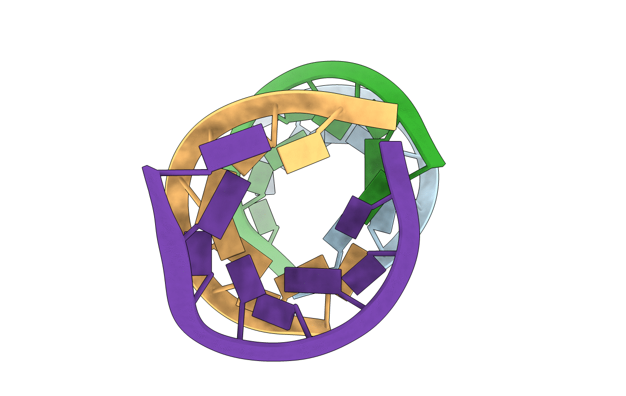
Title:
Crystal structure of the first RNA duplex in L-conformation at 1.9A resolution
Method Details:
Experimental Method:
Resolution:
1.90 Å
R-Value Free:
0.28
R-Value Work:
0.23
R-Value Observed:
0.24
Space Group:
H 3 2