
Deposition Date
2003-10-02
Release Date
2003-11-11
Last Version Date
2024-04-03
Entry Detail
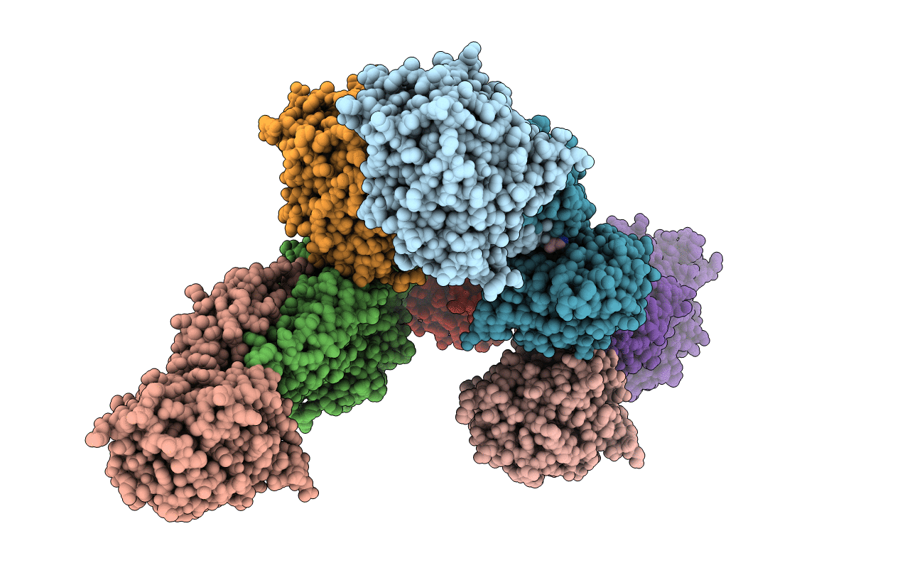
PDB ID:
1R3N
Keywords:
Title:
Crystal structure of beta-alanine synthase from Saccharomyces kluyveri
Biological Source:
Source Organism(s):
Lachancea kluyveri (Taxon ID: 4934)
Expression System(s):
Method Details:
Experimental Method:
Resolution:
2.70 Å
R-Value Free:
0.26
R-Value Work:
0.20
R-Value Observed:
0.21
Space Group:
P 1 21 1
