1R03 image
Deposition Date 2003-09-19
Release Date 2004-06-29
Last Version Date 2023-08-23
Entry Detail
PDB ID:
1R03
Title:
crystal structure of a human mitochondrial ferritin
Biological Source:
Source Organism:
Homo sapiens (Taxon ID: 9606)
Host Organism:
Method Details:
Experimental Method:
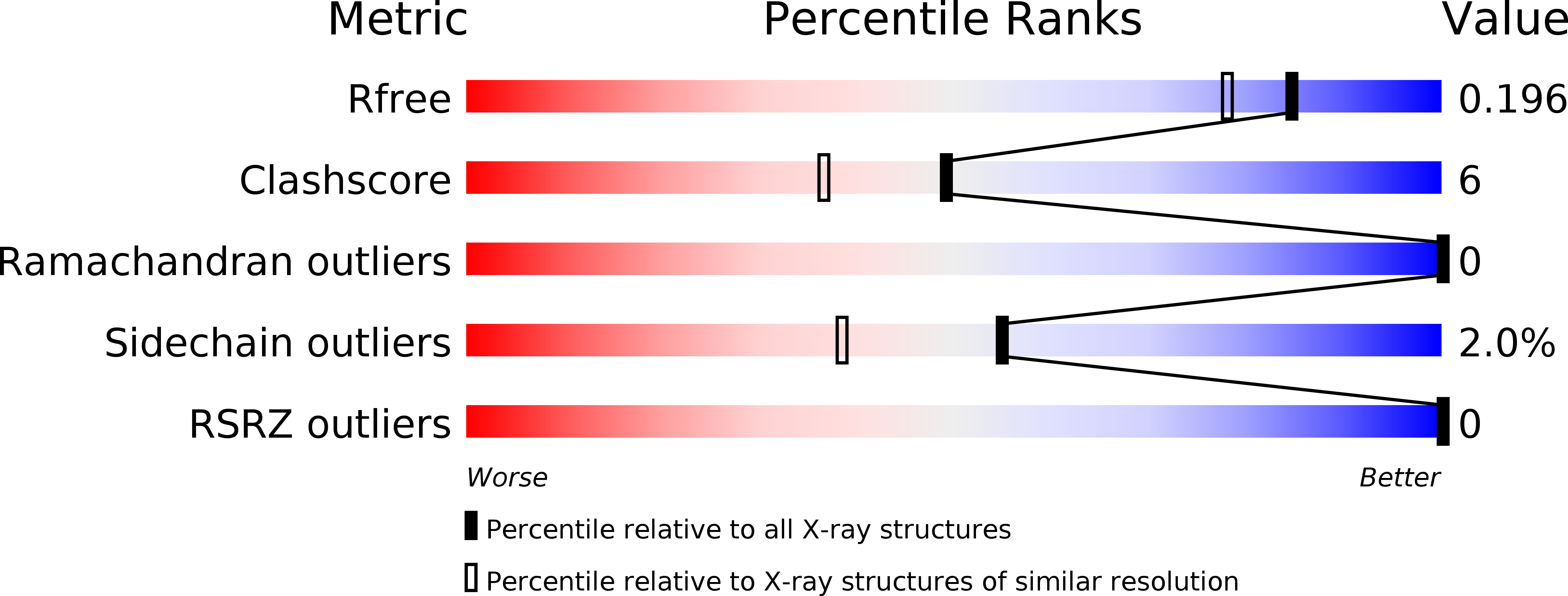
Resolution:
1.70 Å
R-Value Free:
0.2
R-Value Work:
0.17
R-Value Observed:
0.18
Space Group:
F 4 3 2
Macromolecular Entities
Polymer Type:polypeptide(L)
Molecule:mitochondrial ferritin
Gene (Uniprot):FTMT
Chain IDs:A
Chain Length:182
Number of Molecules:1
Biological Source:Homo sapiens
Ligand Molecules
Primary Citation
Crystal Structure and Biochemical Properties of the Human Mitochondrial Ferritin and its Mutant Ser144Ala
J.Mol.Biol. 340 277 293 (2004)
PMID: 15201052 DOI: 10.1016/j.jmb.2004.04.036

Abstact

Mitochondrial ferritin is a recently identified protein precursor encoded by an intronless gene. It is specifically taken up by the mitochondria and processed to a mature protein that assembles into functional ferritin shells. The full mature recombinant protein and its S144A mutant were produced to study structural and functional properties. They yielded high quality crystals from Mg(II) solutions which diffracted up to 1.38 Angstrom resolution. The 3D structures of the two proteins resulted very similar to that of human H-ferritin, to which they have high level of sequence identity (approximately 80%). Metal-binding sites were identified in the native crystals and in those soaked in Mn(II) and Zn(II) solutions. The ferroxidase center binds binuclear iron at the sites A and B, and the structures showed that the A site was always fully occupied by Mg(II), Mn(II) or Zn(II), while the occupancy of the B site was variable. In addition, distinct Mg(II) and Zn(II)-binding sites were found in the 3-fold axes to block the hydrophilic channels. Other metal-binding sites, never observed before in H-ferritin, were found on the cavity surface near the ferroxidase center and near the 4-fold axes. Mitochondrial ferritin showed biochemical properties remarkably similar to those of human H-ferritin, except for the difficulty in renaturing to yield ferritin shells and for a reduced (approximately 41%) rate in ferroxidase activity. This was partially rescued by the substitution of the bulkier Ser144 with Ala, which occurs in H-ferritin. The residue is exposed on a channel that connects the ferroxidase center with the cavity. The finding that the mutation increased both catalytic activity and the occupancy of the B site demonstrated that the channel is functionally important. In conclusion, the present data define the structure of human mitochondrial ferritin and provide new data on the iron pathways within the H-type ferritin shell.

Legend

Protein

Chemical

Disease

Primary Citation of related structures