1QZZ image
Deposition Date 2003-09-19
Release Date 2003-11-25
Last Version Date 2024-02-14
Entry Detail
PDB ID:
1QZZ
Title:
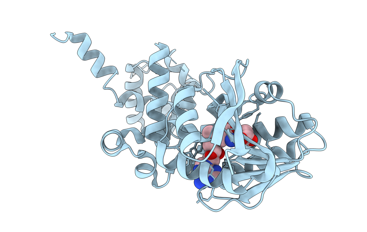
Crystal structure of aclacinomycin-10-hydroxylase (RdmB) in complex with S-adenosyl-L-methionine (SAM)
Biological Source:
Source Organism:
Host Organism:
Method Details:
Experimental Method:
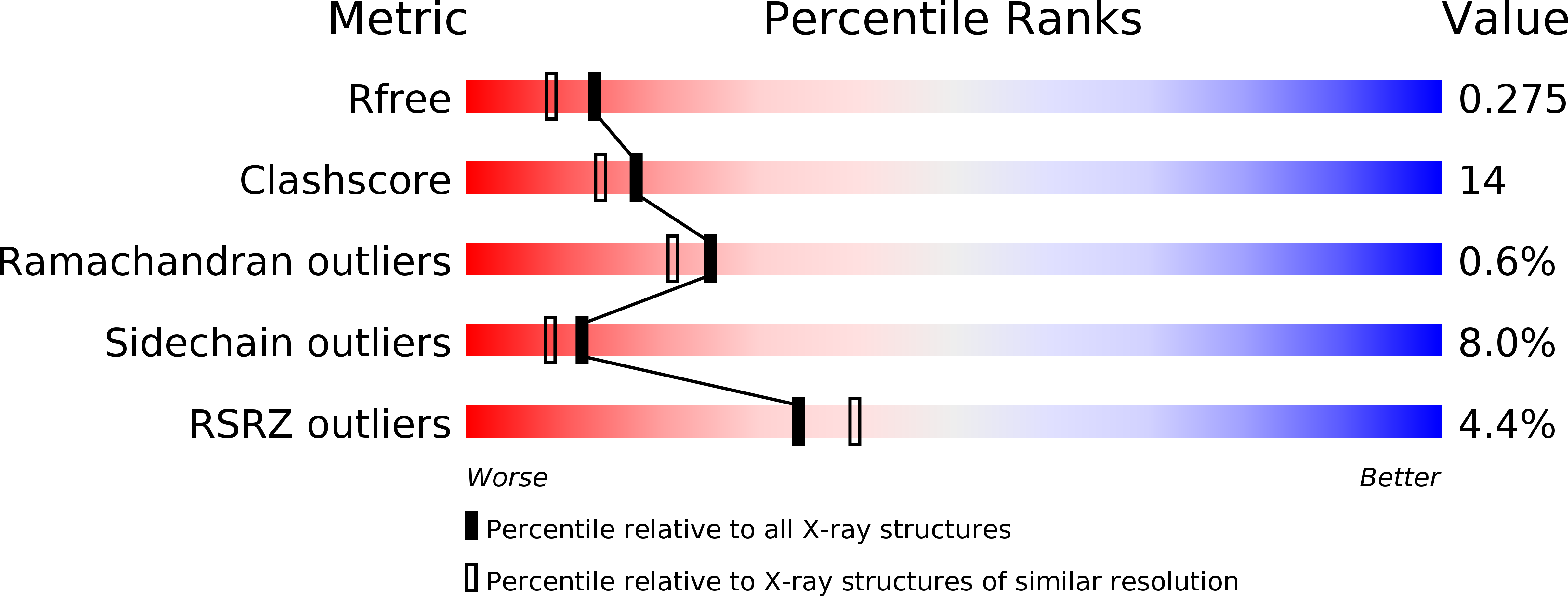
Resolution:
2.10 Å
R-Value Free:
0.27
R-Value Work:
0.20
R-Value Observed:
0.21
Space Group:
C 2 2 21
Macromolecular Entities
Polymer Type:polypeptide(L)
Molecule:aclacinomycin-10-hydroxylase
Gene (Uniprot):rdmB
Chain IDs:A
Chain Length:374
Number of Molecules:1
Biological Source:Streptomyces purpurascens
Primary Citation
Crystal Structure of Aclacinomycin-10-Hydroxylase, a S-Adenosyl-L-Methionine-dependent Methyltransferase Homolog Involved in Anthracycline Biosynthesis in Streptomyces purpurascens.
J.Mol.Biol. 334 269 280 (2003)
PMID: 14607118 DOI: 10.1016/j.jmb.2003.09.061

Abstact

Anthracyclines are aromatic polyketide antibiotics, and several of these compounds are widely used as anti-tumor drugs in chemotherapy. Aclacinomycin-10-hydroxylase (RdmB) is one of the tailoring enzymes that modify the polyketide backbone in the biosynthesis of these metabolites. RdmB, a S-adenosyl-L-methionine-dependent methyltransferase homolog, catalyses the hydroxylation of 15-demethoxy-epsilon-rhodomycin to beta-rhodomycin, one step in rhodomycin biosynthesis in Streptomyces purpurascens. The crystal structure of RdmB, determined by multiwavelength anomalous diffraction to 2.1A resolution, reveals that the enzyme subunit has a fold similar to methyltransferases and binds S-adenosyl-L-methionine. The N-terminal domain, which consists almost exclusively of alpha-helices, is involved in dimerization. The C-terminal domain contains a typical alpha/beta nucleotide-binding fold, which binds S-adenosyl-L-methionine, and several of the residues interacting with the cofactor are conserved in O-methyltransferases. Adjacent to the S-adenosyl-L-methionine molecule there is a large cleft extending to the enzyme surface of sufficient size to bind the substrate. Analysis of the putative substrate-binding pocket suggests that there is no enzymatic group in proximity of the substrate 15-demethoxy-epsilon-rhodomycin, which could assist in proton abstraction and thus facilitate methyl transfer. The lack of a suitably positioned catalytic base might thus be one of the features responsible for the inability of the enzyme to act as a methyltransferase.

Legend

Protein

Chemical

Disease

Primary Citation of related structures