1QZW image
Deposition Date 2003-09-18
Release Date 2003-11-18
Last Version Date 2023-08-23
Entry Detail
PDB ID:
1QZW
Title:
Crystal structure of the complete core of archaeal SRP and implications for inter-domain communication
Biological Source:
Source Organism:
Method Details:
Experimental Method:
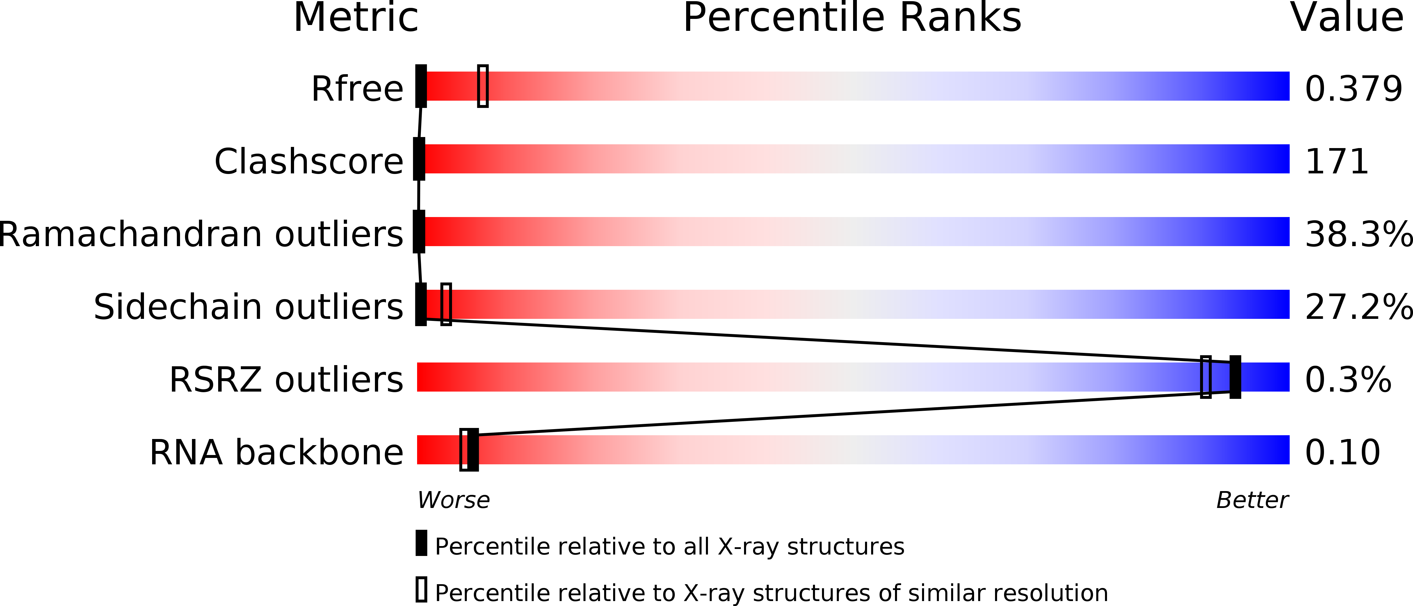
Resolution:
4.10 Å
R-Value Free:
0.38
R-Value Work:
0.33
Space Group:
P 31
Macromolecular Entities
Polymer Type:polypeptide(L)
Molecule:Signal recognition 54 kDa protein
Gene (Uniprot):srp54
Chain IDs:E (auth: A), F (auth: C), G (auth: E), H (auth: G)
Chain Length:440
Number of Molecules:4
Biological Source:Sulfolobus solfataricus
Polymer Type:polyribonucleotide
Molecule:7S RNA
Chain IDs:A (auth: B), B (auth: D), C (auth: F), D (auth: H)
Chain Length:47
Number of Molecules:4
Biological Source:Sulfolobus solfataricus
Modified Residue
Compound ID Chain ID Parent Comp ID Details 2D Image
CCC A C ?
GTP A G GUANOSINE-5'-TRIPHOSPHATE
Ligand Molecules
Primary Citation
Crystal structure of the complete core of archaeal signal recognition particle and implications for interdomain communication
Proc.Natl.Acad.Sci.USA 100 14701 14706 (2003)
PMID: 14657338 DOI: 10.1073/pnas.2436132100

Abstact

Targeting of secretory and membrane proteins by the signal recognition particle (SRP) is evolutionarily conserved, and the multidomain protein SRP54 acts as the key player in SRP-mediated protein transport. Binding of a signal peptide to SRP54 at the ribosome is coordinated with GTP binding and subsequent complex formation with the SRP receptor. Because these functions are localized to distinct domains of SRP54, communication between them is essential. We report the crystal structures of SRP54 from the Archaeon Sulfolobus solfataricus with and without its cognate SRP RNA binding site (helix 8) at 4-A resolution. The two structures show the flexibility of the SRP core and the position of SRP54 relative to the RNA. A long linker helix connects the GTPase (G domain) with the signal peptide binding (M) domain, and a hydrophobic contact between the N and M domains relates the signal peptide binding site to the G domain. Hinge regions are identified in the linker between the G and M domains (292-LGMGD) and in the N-terminal part of the M domain, which allow for structural rearrangements within SRP54 upon signal peptide binding at the ribosome.

Legend

Protein

Chemical

Disease

Primary Citation of related structures