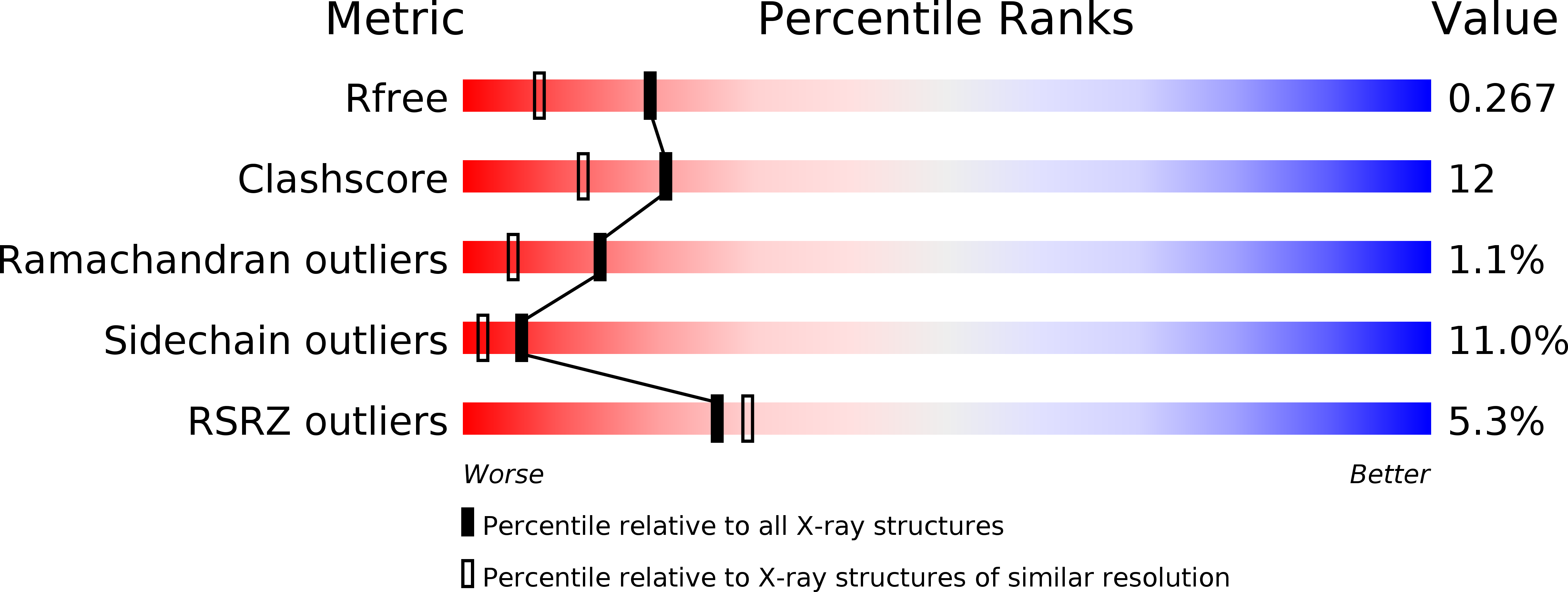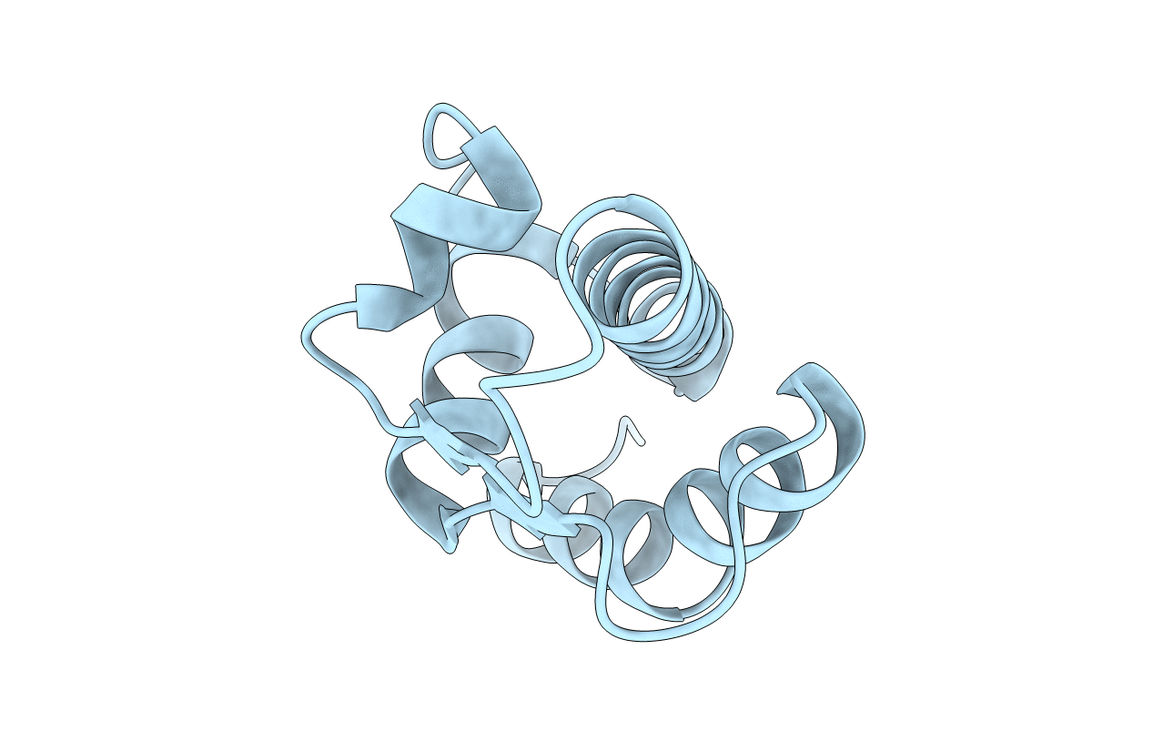
1QZM image
Deposition Date 2003-09-17
Release Date 2004-05-04
Last Version Date 2024-02-14
Entry Detail
PDB ID:
1QZM
Keywords:
Title:
alpha-domain of ATPase
Biological Source:
Source Organism:
Escherichia coli (Taxon ID: 562)
Host Organism:
Method Details:
Experimental Method:
Resolution:
1.90 Å
R-Value Free:
0.28
R-Value Work:
0.17
R-Value Observed:
0.18
Space Group:
P 43 21 2
Macromolecular Entities
Structures with similar UniProt ID
Protein Blast
Polymer Type:polypeptide(L)
Molecule:ATP-dependent protease La
Gene (Uniprot):lon
Chain IDs:A
Chain Length:94
Number of Molecules:1
Biological Source:Escherichia coli
Primary Citation
Crystal structure of the AAA+ alpha domain of E. coli Lon protease at 1.9A resolution.
J.Struct.Biol. 146 113 122 (?)
PMID: 15037242 DOI: 10.1016/j.jsb.2003.09.003

Abstact

The crystal structure of the small, mostly helical alpha domain of the AAA+ module of the Escherichia coli ATP-dependent protease Lon has been solved by single isomorphous replacement combined with anomalous scattering and refined at 1.9A resolution to a crystallographic R factor of 17.9%. This domain, comprising residues 491-584, was obtained by chymotrypsin digestion of the recombinant full-length protease. The alpha domain of Lon contains four alpha helices and two parallel strands and resembles similar domains found in a variety of ATPases and helicases, including the oligomeric proteases HslVU and ClpAP. The highly conserved "sensor-2" Arg residue is located at the beginning of the third helix. Detailed comparison with the structures of 11 similar domains established the putative location of the nucleotide-binding site in this first fragment of Lon for which a crystal structure has become available.

Legend

Protein

Chemical

Disease

Primary Citation of related structures