1QYN image
Deposition Date 2003-09-11
Release Date 2003-12-09
Last Version Date 2023-08-23
Entry Detail
PDB ID:
1QYN
Keywords:
Title:
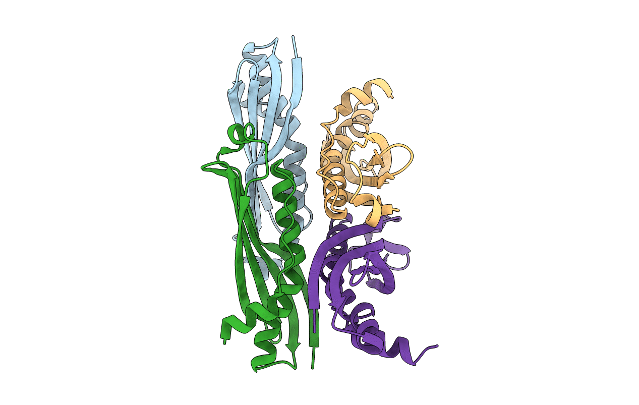
Crystal Structure of SecB from Escherichia coli
Biological Source:
Source Organism:
Escherichia coli (Taxon ID: 562)
Method Details:
Experimental Method:
Resolution:
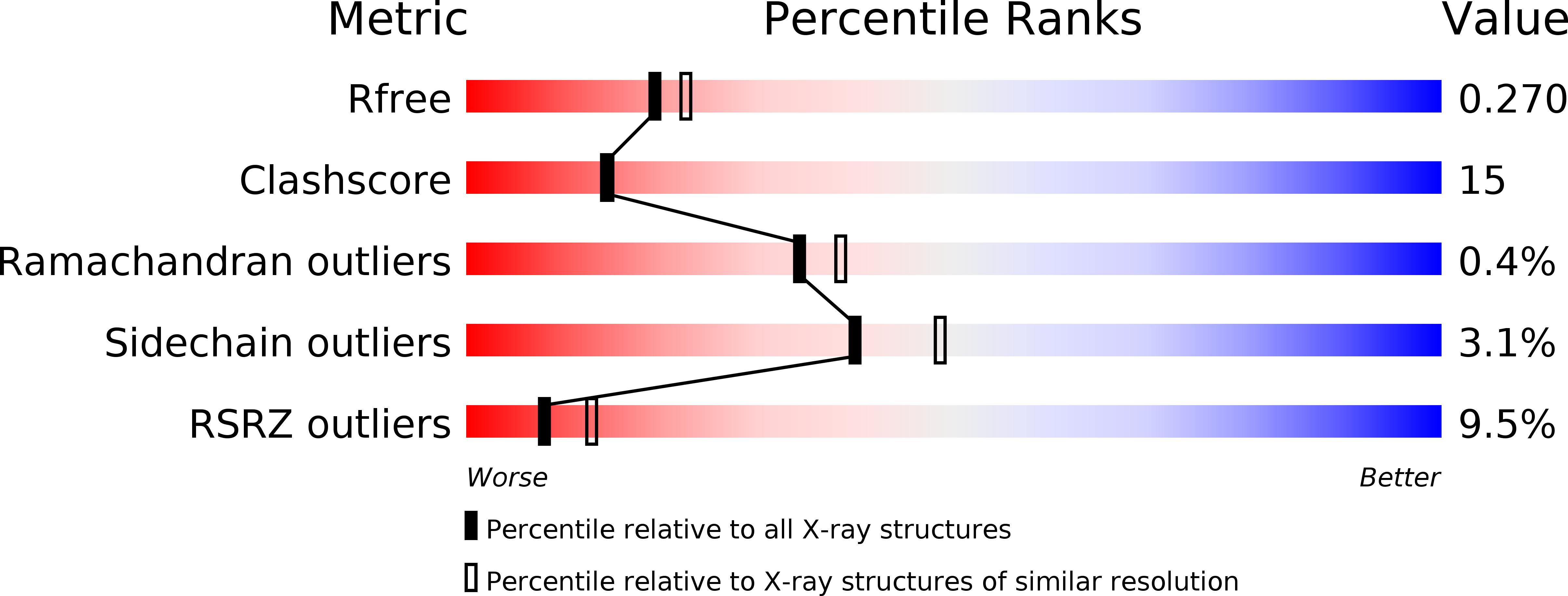
2.35 Å
R-Value Free:
0.27
R-Value Work:
0.23
R-Value Observed:
0.23
Space Group:
P 43 21 2
Macromolecular Entities
Structures with similar UniProt ID
Protein Blast
Polymer Type:polypeptide(L)
Molecule:Protein-export protein secB
Chain IDs:A, B, C, D
Chain Length:153
Number of Molecules:4
Biological Source:Escherichia coli
Primary Citation
Crystal structure of SecB from Escherichia coli
J.Struct.Biol. 144 313 319 (2003)
PMID: 14643199 DOI: 10.1016/j.jsb.2003.09.012

Abstact

The chaperone SecB from Escherichia coli is primarily involved in passing precursor proteins into the Sec system via specific interactions with SecA. The crystal structure of SecB from E. coli has been solved to 2.35 A resolution. The structure shows flexibility in the crossover loop and the helix-connecting loop, regions that have been implicated to be part of the SecB substrate-binding site. Moreover conformational variability of Trp36 is observed as well as different loop conformations for the different monomers. Based on this, we speculate that SecB can regulate the access or extent of its hydrophobic substrate-binding site, by modulating the conformation of the crossover loop and the helix-connecting loop. The structure also clearly explains why the tetrameric equilibrium is shifted towards the dimeric state in the mutant SecBCys76Tyr. The buried cysteine residue is crucial for tight packing, and mutations are likely to disrupt the tetramer formation but not the dimer formation.

Legend

Protein

Chemical

Disease

Primary Citation of related structures