1QYB image
Deposition Date 2003-09-10
Release Date 2004-03-30
Last Version Date 2023-08-23
Entry Detail
PDB ID:
1QYB
Title:
X-ray crystal structure of Desulfovibrio vulgaris rubrerythrin with zinc substituted into the [Fe(SCys)4] site and alternative diiron site structures
Biological Source:
Source Organism:
Method Details:
Experimental Method:
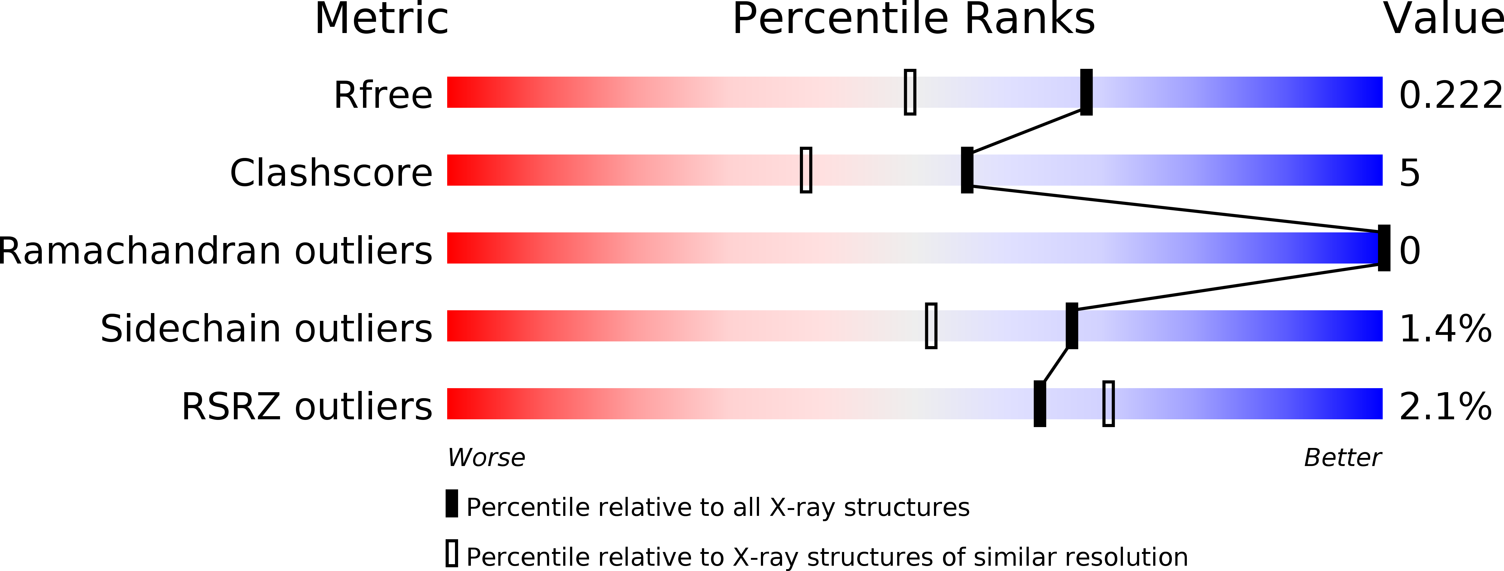
Resolution:
1.75 Å
R-Value Free:
0.21
R-Value Work:
0.18
R-Value Observed:
0.18
Space Group:
I 2 2 2
Macromolecular Entities
Polymer Type:polypeptide(L)
Molecule:Rubrerythrin
Gene (Uniprot):rbr
Chain IDs:A
Chain Length:191
Number of Molecules:1
Biological Source:Desulfovibrio vulgaris subsp. vulgaris
Primary Citation
X-ray Crystal Structure of Desulfovibrio vulgaris Rubrerythrin with Zinc Substituted into the [Fe(SCys)(4)] Site and Alternative Diiron Site Structures.
Biochemistry 43 3204 3213 (2004)
PMID: 15023070 DOI: 10.1021/bi0356193

Abstact

The X-ray crystal structure of recombinant Desulfovibrio vulgaris rubrerythrin (Rbr) that was subjected to metal constitution first with zinc and then iron, yielding ZnS(4)Rbr, is reported. A [Zn(SCys)(4)] site with no iron and a diiron site with no appreciable zinc in ZnS(4)Rbr were confirmed by analysis of the anomalous scattering data. Partial reduction of the diiron site occurred during the synchrotron X-ray irradiation at 95 K, resulting in two different diiron site structures in the ZnS(4)Rbr crystal. These two structures can be classified as containing mixed-valent Fe1(III)(mu-OH(-))(mu-GluCO(2)(-))(2)Fe2(II) and Fe1(II)(mu-GluCO(2)(-))(2)Fe2(III)-OH(-) cores. The data do not show any evidence for alternative positions of the protein or solvent ligands. The iron and ligand positions of the solvent-bridged site are close to those of the diferric site in all-iron Rbr. The diiron site with only the two carboxylato bridges differs by an approximately 2 A shift in the position of Fe1, which changes from six- to four-coordination. The Fe1- - -Fe2 distance (3.6 A) in this latter site is significantly longer than that of the site with the additional solvent bridge (3.4 A) but significantly shorter than that previously reported for the diferrous site (4.0 A) in all-iron Rbr. The apparent redox-induced movement of Fe1 at 95 K in the ZnS(4)Rbr crystal implies an extremely low activation barrier, which is consistent with the rapid (approximately 30 s(-1)) room temperature turnover of the all-iron Rbr during its catalysis of two-electron reduction of hydrogen peroxide. ZnS(4)Rbr does not show peroxidase activity, presumably because the [Zn(SCys)(4)] site, unlike the [Fe(SCys)(4)] site, cannot mediate electron transfer to the diiron site. One or both of the diiron site structures in the cryoreduced ZnS(4)Rbr crystal are likely to represent that (those) of transient mixed-valent diiron site(s) that must occur upon return of the diferric to the diferrous oxidation level during peroxidase turnover.

Legend

Protein

Chemical

Disease

Primary Citation of related structures