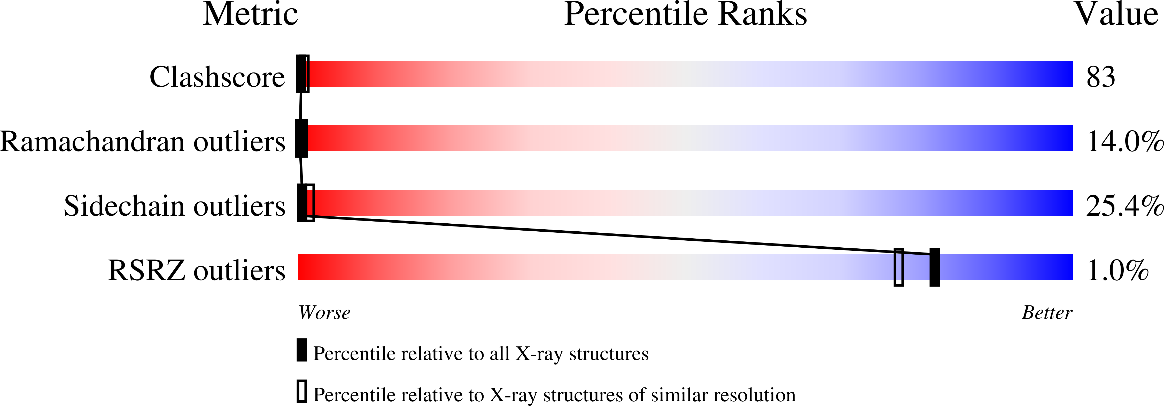
1QXP image
Deposition Date 2003-09-08
Release Date 2004-02-03
Last Version Date 2024-02-14
Entry Detail
PDB ID:
1QXP
Title:
Crystal Structure of a mu-like calpain
Biological Source:
Source Organism:
Rattus norvegicus (Taxon ID: 10116)
Bos taurus (Taxon ID: 9913)
Host Organism:
Method Details:
Experimental Method:
Resolution:
2.80 Å
R-Value Free:
0.31
R-Value Work:
0.22
R-Value Observed:
0.23
Space Group:
P 1 21 1
Macromolecular Entities
Polymer Type:polypeptide(L)
Molecule:mu-like calpain
Gene (Uniprot):Capn1, Capn2
Mutations:C105S
Chain IDs:A, B
Chain Length:900
Number of Molecules:2
Biological Source:Rattus norvegicus, Bos taurus
Primary Citation
Crystal Structure of a mu-like calpain reveals a partially activated conformation with low Ca(2+) requirement
Structure 11 1521 1526 (2003)
PMID: 14656436 DOI: 10.1016/j.str.2003.11.007

Abstact

The two Ca2+-dependent cysteine proteases, micro- and m-calpain, are involved in various Ca2+-linked signal pathways but differ markedly in their Ca2+ requirements for activation. We have determined the structure of a micro-like calpain, which has 85% micro-calpain sequence (the first 48 and the last 62 residues of the large subunit are those from m-calpain) and a low Ca2+ requirement. This construct was used because micro-calpain itself is too poorly expressed. The structure of micro-like calpain is very similar in overall fold to that of m-calpain as expected, but differs significantly in two aspects. In comparison with m-calpain, the catalytic triad residues in micro-like calpain, His and Cys, are much closer together in the absence of Ca2+, and significant portions of the Ca2+ binding EF-hand motifs are disordered and more flexible. These structural differences imply that Ca2+-free micro-calpain may represent a partially activated structure, requiring lower Ca2+ concentration to trigger its activation.

Legend

Protein

Chemical

Disease

Primary Citation of related structures