1QXF image
Deposition Date 2003-09-05
Release Date 2003-09-16
Last Version Date 2024-05-22
Entry Detail
PDB ID:
1QXF
Keywords:
Title:
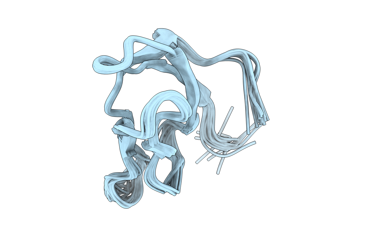
SOLUTION STRUCTURE OF 30S RIBOSOMAL PROTEIN S27E FROM ARCHAEOGLOBUS FULGIDUS: GR2, A NESG TARGET PROTEIN
Biological Source:
Source Organism:
Host Organism:
Method Details:
Experimental Method:
Conformers Calculated:
100
Conformers Submitted:
20
Selection Criteria:
target function
Macromolecular Entities
Polymer Type:polypeptide(L)
Molecule:30S RIBOSOMAL PROTEIN S27E
Gene (Uniprot):rps27e
Chain IDs:A
Chain Length:66
Number of Molecules:1
Biological Source:Archaeoglobus fulgidus
Ligand Molecules
Primary Citation
The NMR solution structure of the 30S ribosomal protein S27e encoded in gene RS27_ARCFU of Archaeoglobus fulgidis reveals a novel protein fold
Protein Sci. 13 1407 1416 (2004)
PMID: 15096641 DOI: 10.1110/ps.03589204

Abstact

The Archaeoglobus fulgidis gene RS27_ARCFU encodes the 30S ribosomal protein S27e. Here, we present the high-quality NMR solution structure of this archaeal protein, which comprises a C4 zinc finger motif of the CX(2)CX(14-16)CX(2)C class. S27e was selected as a target of the Northeast Structural Genomics Consortium (target ID: GR2), and its three-dimensional structure is the first representative of a family of more than 116 homologous proteins occurring in eukaryotic and archaeal cells. As a salient feature of its molecular architecture, S27e exhibits a beta-sandwich consisting of two three-stranded sheets with topology B(decreasing), A(increasing), F(decreasing), and C(increasing), D(decreasing), E(increasing). Due to the uniqueness of the arrangement of the strands, the resulting fold was found to be novel. Residues that are highly conserved among the S27 proteins allowed identification of a structural motif of putative functional importance; a conserved hydrophobic patch may well play a pivotal role for functioning of S27 proteins, be it in archaeal or eukaryotic cells. The structure of human S27, which possesses a 26-residue amino-terminal extension when compared with the archaeal S27e, was modeled on the basis of two structural templates, S27e for the carboxy-terminal core and the amino-terminal segment of the archaeal ribosomal protein L37Ae for the extension. Remarkably, the electrostatic surface properties of archaeal and human proteins are predicted to be entirely different, pointing at either functional variations among archaeal and eukaryotic S27 proteins, or, assuming that the function remained invariant, to a concerted evolutionary change of the surface potential of proteins interacting with S27.

Legend

Protein

Chemical

Disease

Primary Citation of related structures