1QX2 image
Deposition Date 2003-09-04
Release Date 2004-05-25
Last Version Date 2024-11-13
Entry Detail
PDB ID:
1QX2
Title:
X-ray Structure of Calcium-loaded Calbindomodulin (A Calbindin D9k Re-engineered to Undergo a Conformational Opening) at 1.44 A Resolution
Biological Source:
Source Organism:
Bos taurus (Taxon ID: 9913)
Method Details:
Experimental Method:
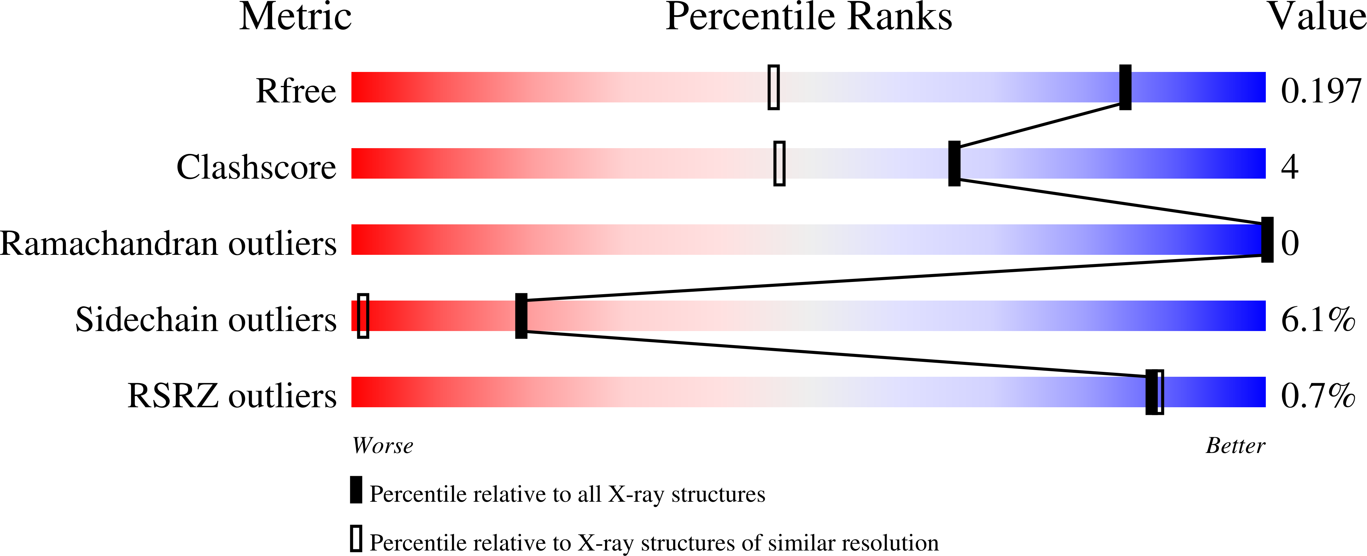
Resolution:
1.44 Å
R-Value Free:
0.19
R-Value Work:
0.15
R-Value Observed:
0.15
Space Group:
C 2 2 21
Macromolecular Entities
Structures with similar UniProt ID
Protein Blast
Polymer Type:polypeptide(L)
Molecule:Vitamin D-dependent calcium-binding protein, intestinal
Gene (Uniprot):S100G
Mutagens:L6I, I9A, K12V, Y13F, L23I, L31V, L32M, E35L, F36G, L49M, F50I, L53V, Q67L, L69M, V70M, P43M
Chain IDs:A, B
Chain Length:76
Number of Molecules:2
Biological Source:Bos taurus
Modified Residue
Compound ID Chain ID Parent Comp ID Details 2D Image
FME A MET N-FORMYLMETHIONINE
Primary Citation
Designing Sequence to Control Protein Function in an EF-Hand Protein
J.Am.Chem.Soc. 126 5990 5998 (2004)
PMID: 15137763 DOI: 10.1021/ja0397456

Abstact

The extent of conformational change that calcium binding induces in EF-hand proteins is a key biochemical property specifying Ca(2+) sensor versus signal modulator function. To understand how differences in amino acid sequence lead to differences in the response to Ca(2+) binding, comparative analyses of sequence and structures, combined with model building, were used to develop hypotheses about which amino acid residues control Ca(2+)-induced conformational changes. These results were used to generate a first design of calbindomodulin (CBM-1), a calbindin D(9k) re-engineered with 15 mutations to respond to Ca(2+) binding with a conformational change similar to that of calmodulin. The gene for CBM-1 was synthesized, and the protein was expressed and purified. Remarkably, this protein did not exhibit any non-native-like molten globule properties despite the large number of mutations and the nonconservative nature of some of them. Ca(2+)-induced changes in CD intensity and in the binding of the hydrophobic probe, ANS, implied that CBM-1 does undergo Ca(2+) sensorlike conformational changes. The X-ray crystal structure of Ca(2+)-CBM-1 determined at 1.44 A resolution reveals the anticipated increase in hydrophobic surface area relative to the wild-type protein. A nascent calmodulin-like hydrophobic docking surface was also found, though it is occluded by the inter-EF-hand loop. The results from this first calbindomodulin design are discussed in terms of progress toward understanding the relationships between amino acid sequence, protein structure, and protein function for EF-hand CaBPs, as well as the additional mutations for the next CBM design.

Legend

Protein

Chemical

Disease

Primary Citation of related structures
Feedback Form
Name
Email
Institute
Feedback